1. Introduction to Structural Organisation
Listen up, future doctors! Before we dive into the nitty-gritty of human physiology, we need to clear up the funda of how living things are built. Think of a multicellular organism like a big Indian wedding. You have individual workers (cells) grouped into teams (tissues), which handle specific stalls (organs), all working together as a management system (organ system) to make sure the biryani doesn't run out! This division of labour is what keeps the body running smoothly.
In this unit, we’ll distinguish between morphology (the external "looks" like the shape of a leaf) and anatomy (the internal "architecture" like the vascular bundles inside a stem). We'll explore how flowering plants are organised into epidermal, ground, and vascular tissue systems, and how their roots, stems, and leaves pull off amazing modifications for survival.
NEET Checkpoint:
What do we call a group of similar cells and their intercellular substances performing a specific function?
(a) Organ
(b) Organ system
(c) Tissue
(d) Organism
✅ Answer: (c) Tissue — This is the basic building block for all complex organs.
NEET Checkpoint:
Does morphology cover internal organs in animals?
(a) Yes, it includes all internal organs
(b) No, it deals with external form and structure
(c) Only reproductive organs
(d) Only skeletal system
✅ Answer: (b) No — In animals, anatomy refers to the morphology of internal organs, while morphology is for external appearance.
Imagine a Pani Puri stall. The cart’s decoration and the size of the puris are its morphology. But the secret mix of the masala and the way the water is stored in different pots? That's the anatomy, yaar!
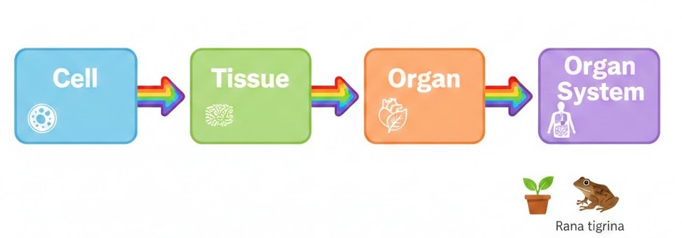
Figure below:
A clear, colourful flowchart showing the biological hierarchy: Cell $\rightarrow$ Tissue $\rightarrow$ Organ $\rightarrow$ Organ System, with a small icon of a plant and a frog ($Rana$ $tigrina$) at the end.
NEET Checkpoint:
Which species of frog is most commonly found in India?
(a) Rana tigrina
(b) Hyla
(c) Bufo
(d) Rana temporaria
✅ Answer: $Rana$ $tigrina$ — Remember this name; it's a frequent NEET guest!
We will also wrap up by looking at a vertebrate model—the frog—to see how its digestive, circulatory, and reproductive systems function. Even if biology feels like a lot to swallow, remember: even a complex system starts with one single cell!
Practice Question:
Which of the following is NOT a part of the three main tissue systems in plants?
(a) Epidermal
(b) Ground
(c) Nervous
(d) Vascular
✅ Answer: (c) Nervous — Plants don't have nerves, though they definitely "feel" the sun!
Key Takeaway
- Hierarchy: Cells $\rightarrow$ Tissues $\rightarrow$ Organs $\rightarrow$ Organ Systems.
- Morphology: Study of external, visible features.
- Anatomy: Study of internal structure and organisation.
2. Plant Tissue Systems: The Three Layers of Life!
Listen up, junior! Understanding plant anatomy is like understanding the layout of a big fat Indian wedding. You’ve got the security at the gate, the guests in the hall, and the catering team running the show. Plants have three main tissue systems based on where they are and what they do: the epidermal, ground, and vascular systems.
The epidermal tissue system is the "skin" of the plant, forming the outermost layer. It includes stomata, which are like tiny windows on leaves for gaseous exchange and transpiration. Think of it like a protective raincoat—most surfaces even have a waxy cuticle to prevent water loss, though the roots skip this to absorb water!
NEET Checkpoint:
What is the shape of guard cells in grasses?
(a) Bean-shaped
(b) Round
(c) Dumb-bell shaped
(d) Heart-shaped
✅ Answer: (c) Dumb-bell shaped — Arre, remember: grass is "buff," so it needs dumb-bells to stay strong!
The ground tissue system is the "filling." It’s basically everything that isn’t the skin or the pipes. It consists of simple tissues like parenchyma, collenchyma, and sclerenchyma. In leaves, this ground tissue is the mesophyll, packed with chloroplasts to cook up food. It’s like the aloo filling in a samosa—it’s where the real substance is!
NEET Checkpoint:
Are root hairs unicellular or multicellular?
(a) Multicellular
(b) Unicellular
(c) Both
(d) None
✅ Answer: (b) Unicellular — They are lonely explorers, while stem trichomes are usually the "party crowd" (multicellular).
Finally, the vascular tissue system consists of the "delivery pipes": xylem and phloem. Together, they form vascular bundles. If they have cambium between them (in dicots), they are "open" for business (secondary growth); otherwise, they are "closed".
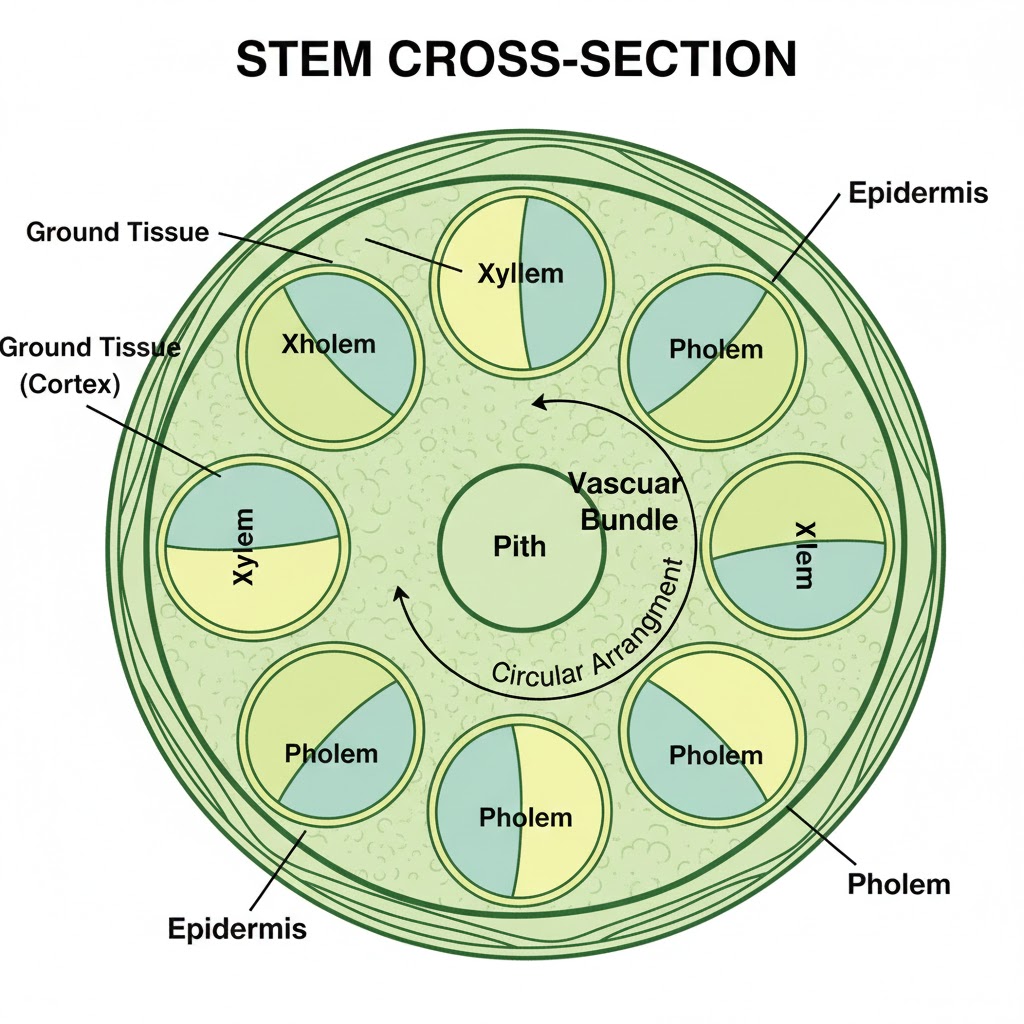
Figure below:
A clear diagram showing the cross-section of a stem with the outermost epidermal layer, the thick ground tissue (cortex), and the circular arrangement of vascular bundles containing xylem and phloem.
NEET Checkpoint:
Which tissue system is the mesophyll part of?
(a) Dermal tissue system
(b) Vascular tissue system
(c) Ground tissue system
(d) Support tissue system
✅ Answer: (c) Ground tissue system — It’s the functional "middle" of the leaf.
Practice Question:
In which organ would you find a radial vascular bundle arrangement?
(a) Dicot stem
(b) Monocot leaf
(c) Roots
(d) Flower petals
✅ Answer: (c) Roots — In roots, xylem and phloem are on different radii.
Key Takeaway
- Epidermal: Outer protection, stomata, and trichomes.
- Ground: Main bulk; contains parenchyma, collenchyma, and sclerenchyma.
- Vascular: Conducting tissues; Xylem + Phloem = Vascular Bundle.
3. Morphology and Modifications of the Root
Hey future doc! Let's dig into the roots—literally. Don't worry, it’s not a "deep" struggle, it’s actually quite a simple funda! In most dicots, the radicle elongates to form the primary root, which branches out into secondary and tertiary roots. This whole setup is the tap root system, like you see in a mustard plant.
NEET Checkpoint:
Which plant has a root system originating from the base of the stem?
(a) Mustard
(b) Wheat
(c) Banyan
(d) Monstera
✅ Answer: (b) Wheat — This is the fibrous root system typical of monocots where the primary root is short-lived.
Then we have the "rebels" called adventitious roots. These arise from parts other than the radicle, like in grass or the massive banyan tree. Think of tap roots as a solid cricket stump planted deep, while fibrous roots are like the messy bundle of wires you see behind a local rickshaw! The root’s job isn't just "drinking" water and minerals; it also provides anchorage, stores food, and even synthesises plant growth regulators.
NEET Checkpoint:
Roots of Monstera are an example of:
(a) Tap roots
(b) Fibrous roots
(c) Adventitious roots
(d) Primary roots
✅ Answer: (c) Adventitious roots — Arre, remember they arise from parts other than the radicle!
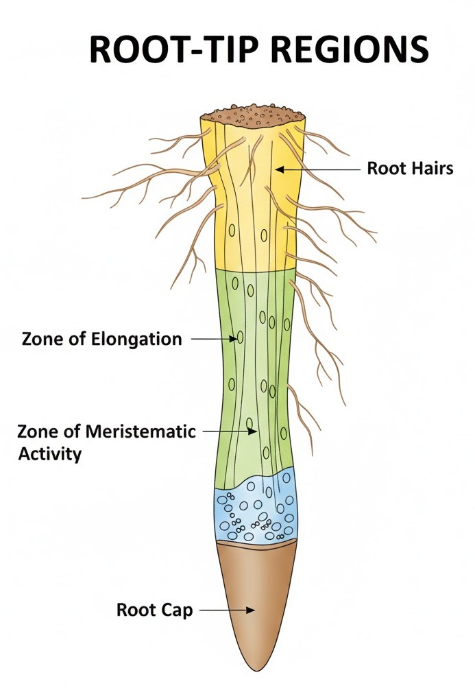
Looking at the root tip, it’s protected by a thimble-like root cap. Just above it is the region of meristematic activity where cells are tiny and divide like crazy. Next is the region of elongation, making the root grow long, followed by the region of maturation where you find those delicate root hairs.
Figure below:
A clear diagram showing the regions of the root-tip, including the root cap, the zone of meristematic activity, the zone of elongation, and the zone of maturation with fine root hairs protruding.
NEET Checkpoint:
Which region is responsible for the growth of the root in length?
(a) Root cap
(b) Maturation
(c) Elongation
(d) Meristematic
✅ Answer: (c) Elongation — The name says it all, yaar! These cells enlarge to increase length.
Practice Questions:
1. Root hairs develop from the region of:
(a) Elongation
(b) Maturation
(c) Meristematic activity
(d) Root cap
✅ Answer: (b) Root hairs are epidermal elongations found in the maturation zone.
2. The main functions of the root system include:
(a) Absorption of water
(b) Providing anchorage
(c) Storing reserve food
(d) All of these
✅ Answer: (d) As per the sources, roots handle absorption, anchorage, storage, and regulator synthesis.
Key Takeaway
- Tap Root: Dicot (Mustard), from radicle.
- Fibrous Root: Monocot (Wheat), from stem base.
- Adventitious Root: (Banyan/Grass), non-radicle origin.
- Zones: Root Cap $\rightarrow$ Meristematic $\rightarrow$ Elongation $\rightarrow$ Maturation (Root Hairs).
4. Anatomy of Dicot and Monocot Roots
Hey junior! Let’s get to the 'root' of the matter. Analyzing the internal structure (anatomy) of roots is actually quite a chill funda once you see the patterns.
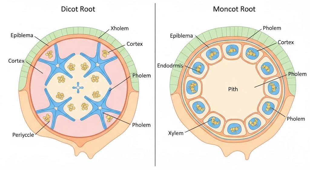
In a Dicot root, like our friend the sunflower, the outer layer is the epiblema, which sports unicellular root hairs. Below that is a massive cortex leading to the endodermis. These endodermal cells have Casparian strips made of waxy suberin—think of it as natural waterproof tape!. You'll only find a few ($2$ to $4$) xylem and phloem patches here.
NEET Checkpoint:
What is the waxy material in Casparian strips?
(a) Cutin
(b) Suberin
(c) Lignin
(d) Cellulose
✅ Answer: (b) Suberin — It is a water-impermeable material deposited on the tangential and radial walls of endodermal cells.
Now, Monocot roots are the "extra" siblings. While they have the same basic layers, they are polyarch, meaning they typically have more than $6$ xylem bundles! Their pith is also large and well-developed, whereas in dicots, it’s tiny or even invisible.
Relatable Example: Think of a Pani Puri plate. A Dicot root is like a standard plate with just $2-4$ puris (xylem patches). A Monocot root is the "Mega Party Pack" with more than $6$ (polyarch) puris arranged in a circle!.
NEET Checkpoint:
Which root type has a large, well-developed pith?
(a) Taproot
(b) Fibrous root
(c) Dicotyledonous root
(d) Monocotyledonous root
✅ Answer: (d) Monocotyledonous root — Unlike dicots, monocot roots need that central space.
One major difference: Dicots have "career growth" plans, meaning they undergo secondary growth to get thicker. Monocots? They are happy as they are and do not undergo any secondary growth.
NEET Checkpoint:
Where do lateral roots originate?
(a) Epidermis
(b) Cortex
(c) Pericycle
(d) Endodermis
✅ Answer: (c) Pericycle — The initiation of lateral roots happens in these thick-walled parenchymatous cells.
Figure below:
A comparative transverse section (T.S.) diagram showing a Dicot root with 4 xylem patches and a small pith vs. a Monocot root with 8+ xylem patches (polyarch) and a large central pith. Labels: Epiblema, Cortex, Endodermis, Pericycle, Phloem, and Xylem.
Practice Question:
A student observes a root section under a microscope and finds $8$ xylem bundles and a conspicuous pith. This specimen is:
(a) Dicot root
(b) Monocot root
(c) Dicot stem
(d) Monocot stem
✅ Answer: (b) — Monocot roots are polyarch (more than $6$ xylem bundles) and have a large pith.
Key Takeaway
- Dicot Root: $2-4$ xylem patches, small pith, secondary growth present.
- Monocot Root: $>6$ xylem patches (polyarch), large pith, secondary growth absent.
5. Morphology and Functions of the Stem
Arre junior, don't get confused between a root and a stem! While the root is a "down-to-earth" guy, the stem is the ascending superstar of the plant axis. It develops directly from the plumule of the embryo during germination. Think of the stem as the main chassis of a scooter; it’s the structural frame that holds the "passengers"—the branches, leaves, flowers, and fruits.
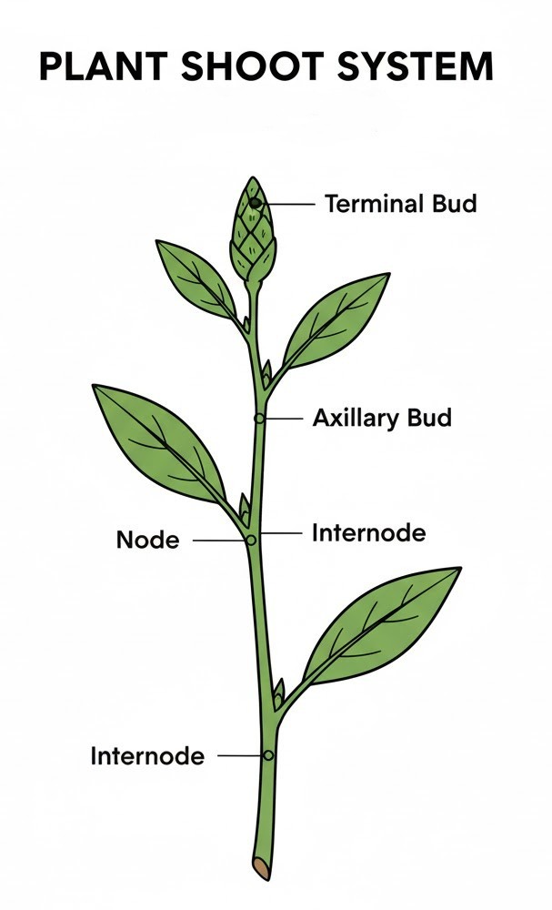
The real funda to identify a stem is looking for nodes and internodes. Nodes are like the busy bus stops where leaves "board" the stem, while the internodes are the long stretches of road between those stops. You’ll also find buds here, which can be terminal (at the tip) or axillary (in the leaf's "armpit"). While young stems are often green and do a bit of photosynthesis, they usually grow up to be woody and dark brown.
NEET Checkpoint:
From which part of the embryo does the stem originate?
(a) Radicle
(b) Plumule
(c) Endosperm
(d) Cotyledon
✅ Answer: (a) Plumule — Remember, Radicle makes the Root, and Plumule makes the "Post-ground" shoot!
Beyond just standing tall, the stem is the plant's plumbing system. It conducts water, minerals, and photosynthates (food) between the roots and leaves. Some stems are even multi-taskers! They can modify themselves to store food (like a hidden tiffin), provide support, offer protection, or even help in vegetative propagation.
Figure below:
A clear, labelled diagram of a shoot system highlighting the terminal bud, axillary bud, a node, and an internode on a vertical stem axis.
NEET Checkpoint:
Which feature helps distinguish a stem from a root?
(a) Presence of root hairs
(b) Presence of nodes and internodes
(c) Absence of vascular tissue
(d) Presence of apical meristem
✅ Answer: (b) Presence of nodes and internodes — Roots don't have these "junctions", yaar!
NEET Checkpoint:
Stems are always the "green" part of the plant.
(a) True
(b) False
✅ Answer: (b) False — They are generally green when young but often become woody and dark brown as they mature.
Practice Question:
Which of the following is a primary function of the stem?
(a) Absorption of water from soil
(b) Anchorage to the plant
(c) Spreading out branches bearing leaves
(d) Synthesis of plant growth regulators
✅ Answer: (c) — Absorption and anchorage are the root's department; the stem's main job is spreading the canopy for sunlight.
Key Takeaway
- Origin: Developed from the plumule.
- Structure: Features nodes, internodes, and buds (terminal/axillary).
- Primary Functions: Conduction of water/minerals and spreading branches.
- Secondary Functions: Storage, support, protection, and propagation.
6. Anatomy of Dicot and Monocot Stems
Hey there, future doctor! Ready to slice through the internal secrets of stems? Don't stress, yaar—it's easier than it looks! Let's clear up this funda.
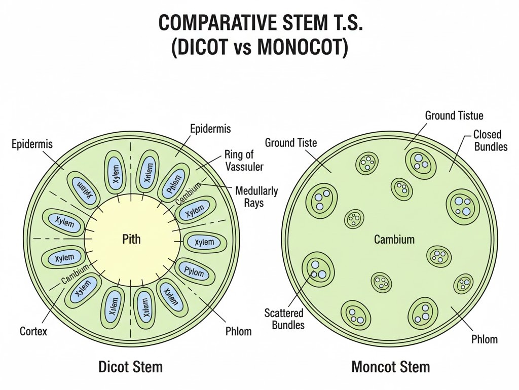
In a Dicotyledonous stem, everything is very "VIP" and organized. The vascular bundles are arranged in a distinct ring, which is the hallmark of dicots. Imagine a group of friends sitting in a perfect circle at a dhaba—that’s your dicot arrangement! These bundles are conjoint and open, meaning they have a cambium between the xylem and phloem, giving them the "open" permission for secondary growth.
NEET Checkpoint:
Which layer is known as the 'starch sheath' in dicot stems?
(a) Pericycle
(b) Endodermis
(c) Hypodermis
(d) Epidermis
✅ Answer: (b) Endodermis — Because these cells are super rich in starch grains!
Now, Monocot stems are the rebels. Their vascular bundles are scattered throughout the large, parenchymatous ground tissue. It’s like a crowded Mumbai local train where everyone is standing wherever they find space! These bundles are conjoint and closed (no cambium, so no secondary growth) and are protected by a tough sclerenchymatous bundle sheath.
NEET Checkpoint:
In which stem is phloem parenchyma absent?
(a) Dicot
(b) Monocot
(c) Both
(d) None
✅ Answer: (b) Monocot — It's a key anatomical difference you must remember!
Notice this: dicots have a distinct pith in the centre and medullary rays between bundles, while monocots just have a massive "ground tissue". Even the hypodermis differs—dicots use collenchyma for flexible strength, while monocots use sclerenchyma.
NEET Checkpoint:
Peripheral vascular bundles in monocot stems are generally:
(a) Larger than central
(b) Smaller than central
(c) Same size
(d) Absent
✅ Answer: (b) Smaller than central — Notice this pattern in the diagram!
Figure below:
A clear diagram comparing the T.S. of a Dicot stem showing a ring of open vascular bundles, pith, and medullary rays, versus a Monocot stem showing scattered closed vascular bundles and a large ground tissue.
Practice Question:
A plant showing conjoint, closed vascular bundles and water-containing cavities is a:
(a) Sunflower
(b) Mustard
(c) Maize
(d) Peepal
✅ Answer: (c) Maize — Closed bundles and water cavities are classic monocot features!
Key Takeaway
- Dicot Stem: Ring arrangement, Open bundles (cambium), distinct pith, collenchymatous hypodermis.
- Monocot Stem: Scattered, Closed bundles, bundle sheath, sclerenchymatous hypodermis, no phloem parenchyma.
7. Morphology and Venation of the Leaf
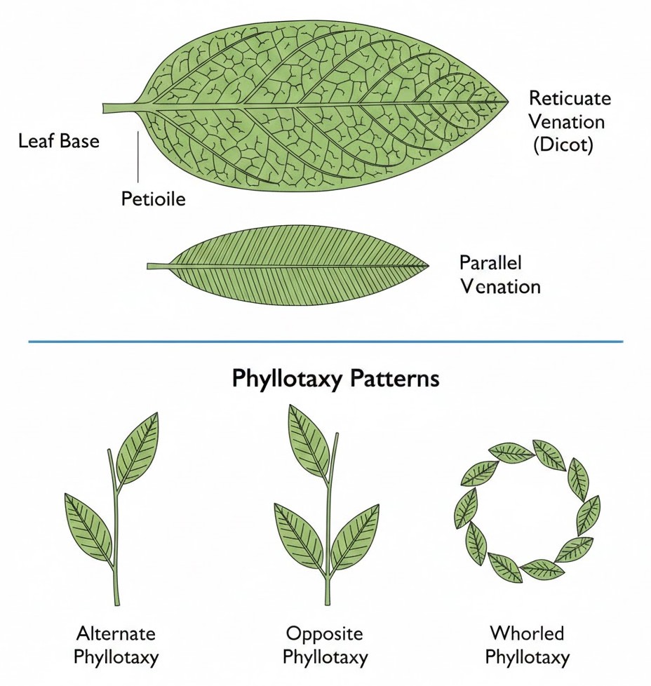
Hey there, future doctor! Let’s talk about the green "food factories" of the plant—the leaves. A leaf is a lateral, flattened structure that develops at the node of a stem and is the most important organ for photosynthesis. A typical leaf has three main parts: the leaf base, the petiole, and the lamina.
Think of the petiole as a flexible stalk that lets the leaf "flutter" in the wind to stay cool—just like you waving a hand-fan during a summer power cut in the hostel! In some leguminous plants, the leaf base becomes swollen and is called the pulvinus. The flat green part is the lamina, which has a prominent middle vein called the midrib.
NEET Checkpoint:
What is the name of the swollen leaf base found in some legumes?
(a) Petiole
(b) Pulvinus
(c) Stipule
(d) Petiolule
✅ Answer: (b) Pulvinus — It's a "swollen" base, not a puffy snack!
Next up is venation, which is the arrangement of veins in the lamina. If the veins form a messy network (like the complex routes of a local rickshaw puller), it’s reticulate venation, common in dicots. If they run parallel, like the straight tracks of the Metro, it’s parallel venation, typical for monocots.
NEET Checkpoint:
Which venation is a characteristic of most monocotyledonous plants?
(a) Reticulate venation
(b) Parallel venation
(c) Pinnate venation
(d) Palmate venation
✅ Answer: (b) Parallel venation — Straight lines, no drama!
Finally, phyllotaxy is the plant's seating chart—how leaves are arranged on the stem. It can be alternate (one leaf per node, like China rose), opposite (two leaves per node, like Guava), or whorled (more than two leaves forming a circle, like Alstonia).
NEET Checkpoint:
In which plant would you find whorled phyllotaxy?
(a) Guava
(b) China rose
(c) Alstonia
(d) Mustard
✅ Answer: (c) Alstonia — It’s the one where the leaves gather in a group circle!
Figure below:
A clear diagram showing the parts of a typical leaf including the leaf base, petiole, and lamina; also showing reticulate venation in a dicot leaf versus parallel venation in a monocot leaf, followed by clear examples of alternate, opposite, and whorled phyllotaxy.
Practice Question:
A single leaf arises at each node in an alternate manner in:
(a) Calotropis
(b) Guava
(c) China rose
(d) Alstonia
✅ Answer: (c) China rose — In mustard, sunflower, and China rose, the leaves follow the "one-at-a-time" rule.
Key Takeaway
• Leaf Parts: Base (can have stipules/pulvinus), Petiole, and Lamina.
• Venation: Reticulate in dicots; Parallel in monocots.
• Phyllotaxy: Alternate (China rose), Opposite (Guava), Whorled (Alstonia).
8. Anatomy of Dorsiventral and Isobilateral Leaves
Namaste, junior! Ready to dive into the internal world of leaves? Don't stress, yaar—it's easier than it looks! Let's clear up this funda about how Dicots and Monocots keep their "kitchens" (leaves) organized internally.
The Dorsiventral (Dicot) Leaf
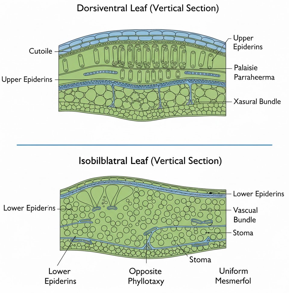
Think of a dorsiventral leaf as a fancy Mumbai duplex apartment. It has a clear "top" (adaxial epidermis) and "bottom" (abaxial epidermis). The top floor is usually shy and has fewer or no stomata, while the bottom floor is the main entrance with most of the stomata for gaseous exchange.
The middle part, the mesophyll, is where the magic happens. It's differentiated into two teams:
1. Palisade parenchyma: These cells are like students in a crowded coaching class—standing tall, packed vertically, and parallel to each other.
2. Spongy parenchyma: These guys are chilled out! They are oval or round and loosely arranged with huge air cavities to let the leaf "breathe".
NEET Checkpoint:
In a dicot leaf, where would you find the most stomata?
(a) Adaxial epidermis
(b) Abaxial epidermis
(c) Palisade parenchyma
(d) Midrib
✅ Answer: (b) Abaxial epidermis — It helps prevent excessive water loss from direct sunlight!
The Isobilateral (Monocot) Leaf
Now, the isobilateral leaf is way more democratic! Stomata are present on both surfaces, and the mesophyll refuses to take sides—it is not differentiated into palisade and spongy layers.
In grasses, there is a super cool feature: bulliform cells. Imagine a local rickshaw puller pulling down the side covers during a dusty storm—these large, empty cells on the adaxial epidermis do exactly that! When they lose water and go flaccid, they make the leaves curl inwards to minimize water loss.
NEET Checkpoint:
What is the function of bulliform cells in grasses?
(a) Photosynthesis
(b) To minimize water loss
(c) Water absorption
(d) Transport of nutrients
✅ Answer: (b) To minimize water loss — They make the leaves curl when the plant is under water stress!
Figure below:
A detailed vertical section diagram of a dorsiventral leaf showing the packed palisade parenchyma at the top and loose spongy parenchyma with air cavities at the bottom, and an isobilateral leaf showing uniform mesophyll and large bulliform cells in the upper epidermis.
NEET Checkpoint:
Which type of venation leads to near-similar sizes of vascular bundles in monocot leaves?
(a) Reticulate venation
(b) Parallel venation
(c) Pinnate venation
(d) Palmate venation
✅ Answer: (b) Parallel venation — Except for the main veins, most bundles look the same size in cross-section!
Practice Question:
Which of the following is characteristic of a monocot leaf?
(a) Differentiated mesophyll
(b) Stomata only on the abaxial surface
(c) Bulliform cells in grasses
(d) Reticulate venation
✅ Answer: (c) Bulliform cells in grasses — These are specialized cells for handling water stress!
Key Takeaway:
- Dorsiventral (Dicot): Different top/bottom; differentiated mesophyll (Palisade + Spongy).
- Isobilateral (Monocot): Same on both sides; undifferentiated mesophyll; contains bulliform cells in grasses.
9. Inflorescence: Cymose and Racemose
Ever wondered why some plants look like a tall tower of flowers while others look like a flat bouquet? It’s all about the inflorescence, which is the technical way of describing the arrangement of flowers on the floral axis. Think of it like the seating plan at a big desi wedding—where everyone sits depends on the "axis" of the family!.
NEET Checkpoint:
What is the term for the arrangement of flowers on the floral axis?
(a) Phyllotaxy
(b) Venation
(c) Inflorescence
(d) Aestivation
✅ Answer: (c) Inflorescence — Simple funda: leaves have phyllotaxy, flowers have inflorescence.
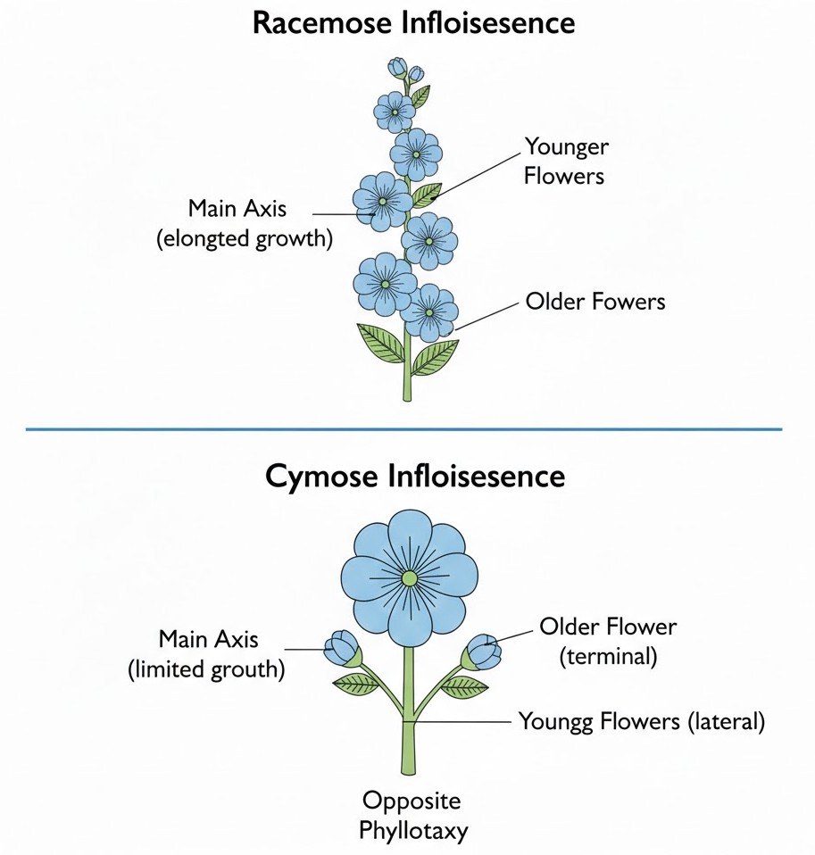
In the Racemose type, the main axis is a total overachiever—it continues to grow indefinitely. The flowers are borne laterally in an acropetal succession. This means the youngest flowers are at the top and the older ones are at the base. It’s exactly like a queue at a Pani Puri stall where the line just keeps growing longer as new people join at the end!.
NEET Checkpoint:
In a racemose inflorescence, the flowers are arranged in:
(a) Basipetal order
(b) Acropetal succession
(c) Whorls
(d) Randomly
✅ Answer: (b) Acropetal succession — The main axis never stops growing, and new flowers keep appearing at the apex.
In contrast, the Cymose inflorescence is a bit more "chilled." The main axis terminates in a flower, which means its growth is limited. Here, the flowers are arranged in a basipetal order. The oldest flower is at the top, and the younger ones are below.
Figure below:
A clear comparison diagram showing two floral axes. The first (Racemose) shows an elongated main axis with younger flowers at the top. The second (Cymose) shows the main axis ending in a large, older flower with younger flowers developing on lower lateral branches.
NEET Checkpoint:
Which type of inflorescence shows limited growth of the main axis?
(a) Racemose
(b) Cymose
(c) Reticulate
(d) Parallel
✅ Answer: (b) Cymose — Because the tip becomes a flower, the axis says "bas, aur nahi!" (no more!).
Practice Question:
A plant has a main axis that ends in a flower. Which type of inflorescence is this?
(a) Racemose
(b) Cymose
(c) Solitary
(d) None of these
✅ Answer: (b) Cymose — The termination of the axis into a flower is the signature move of cymose.
Key Takeaway
- Racemose: Main axis continues to grow; flowers in acropetal succession (youngest at top).
- Cymose: Main axis ends in a flower (limited growth); flowers in basipetal order (youngest at bottom).
10. The Flower: Structure and Whorls
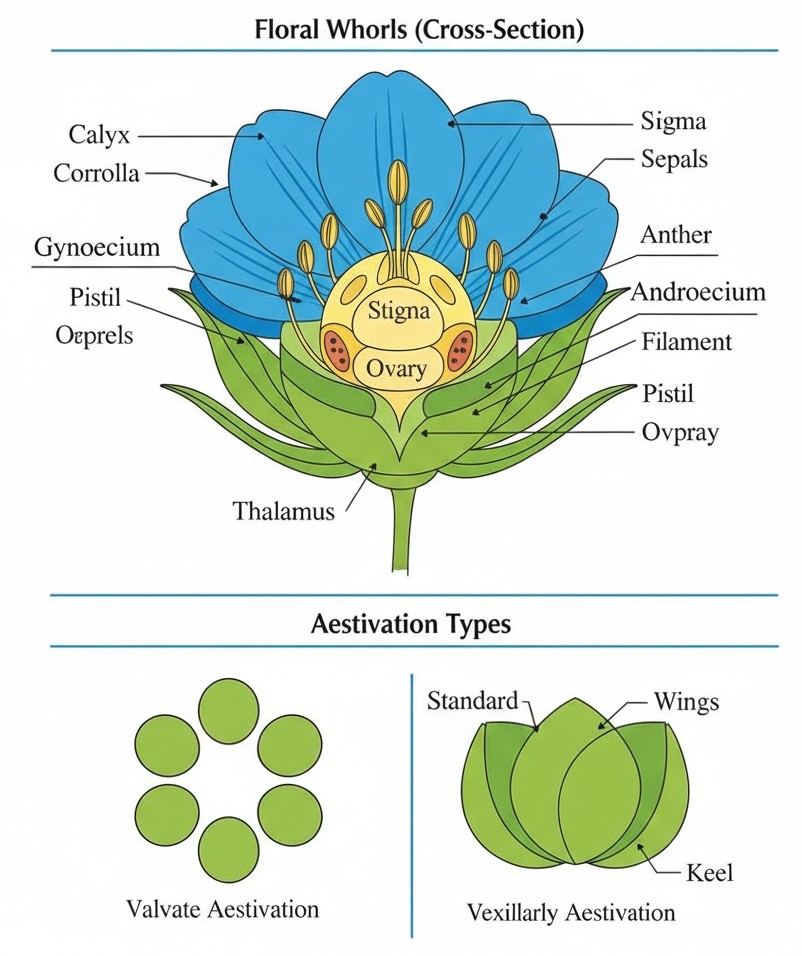
Namaste, future doctors! Let’s talk about the most beautiful part of a plant—the flower. In biology, it’s not just for bouquets; it is the reproductive unit of angiosperms. Think of the flower as a four-story building sitting on a swollen base called the thalamus or receptacle.
A typical flower has four whorls. The outer two are the "security and decor" team: the Calyx (sepals) and Corolla (petals). The inner two are the real VIPS: the Androecium (male stamens) and Gynoecium (female carpels).
NEET Checkpoint:
What do you call it when the calyx and corolla are not distinct?
(a) Polypetalous
(b) Perianth
(c) Gamopetalous
(d) Monocotyledonous
✅ Answer: (b) Perianth — As seen in Lily, it’s like the sepals and petals decided to wear matching uniforms!
Now, check out Aestivation—the way sepals or petals are arranged in a bud. It’s like arranging chairs for a shaadi (wedding). If they just touch, it's Valvate (Calotropis). If they overlap like a stack of parathas, it’s Twisted (China rose). If it’s a mess with no direction, it’s Imbricate (Gulmohur).
NEET Checkpoint:
Which aestivation is found in Pea flowers?
(a) Valvate
(b) Twisted
(c) Vexillary
(d) Imbricate
✅ Answer: (c) Vexillary — It has one large 'standard', two 'wings', and two fused 'keels'. It’s the VIP seating of the plant world!
Lastly, we have Placentation—the "seating arrangement" of ovules inside the ovary. In Axile (lemon), ovules are at the centre, while in Marginal (pea), they form a ridge.
NEET Checkpoint:
Where do you find Basal placentation?
(a) Sunflower
(b) Mustard
(c) China rose
(d) Argemone
✅ Answer: (a) Sunflower — Here, a single ovule is attached at the base, keeping it simple!
Figure below:
A clear diagram showing a cross-section of a flower with four distinct whorls (calyx, corolla, androecium, and gynoecium) arranged on a swollen thalamus, along with sub-diagrams for Valvate and Vexillary aestivation.
Practice Question:
In which plant would you find a syncarpous ovary (fused carpels)?
(a) Rose
(b) Lotus
(c) Mustard
(d) Tomato
✅ Answer: (d) Tomato — 'Syn' means together; these carpels are fused like a tight-knit family!
Key Takeaway
- Four Whorls: Calyx, Corolla, Androecium, Gynoecium.
- Aestivation: Arrangement of sepals/petals (Valvate, Twisted, Imbricate, Vexillary).
- Placentation: Ovule arrangement (Marginal, Axile, Parietal, Free central, Basal).
11. Structure of Fruits and Seeds
Listen up, junior! We’ve reached the "sweet" part of the chapter—literally. After all that flowering drama, the plant finally settles down to produce fruits and seeds. Think of the fruit as the "prize" and the seed as the "future" packed in a tiny box.
A fruit is simply a ripened ovary developed after fertilisation. If a fruit forms without this "meeting," we call it parthenocarpic (like a lonely banana). In fruits like mango and coconut, known as drupes, the ovary is superior and monocarpellary.
NEET Checkpoint:
A fruit formed without fertilisation is called:
(a) Drupe
(b) Parthenocarpic
(c) Dicot
(d) Endospermous
✅ Answer: (b) Parthenocarpic — Easy funda, yaar! No fertilisation = Parthenocarpic.
In a mango, the pericarp is well-differentiated. You have the thin epicarp (skin), the fleshy mesocarp (the juicy part we gobble up), and the stony endocarp (the hard part inside). In a coconut, the mesocarp is actually fibrous, not fleshy! Imagine peeling a coconut at a temple—that husk is just the fibrous mesocarp.
NEET Checkpoint:
Which layer is edible in a mango?
(a) Endocarp
(b) Epicarp
(c) Mesocarp
(d) Perisperm
✅ Answer: (c) Mesocarp — It’s the middle fleshy layer that makes mangoes the king of fruits!
Now, let's talk seeds. They develop from ovules. Dicot seeds (like your favorite chana or pea) have two cotyledons. Most are non-endospermous, meaning they consume the endosperm before they mature, though castor is an exception that keeps it!
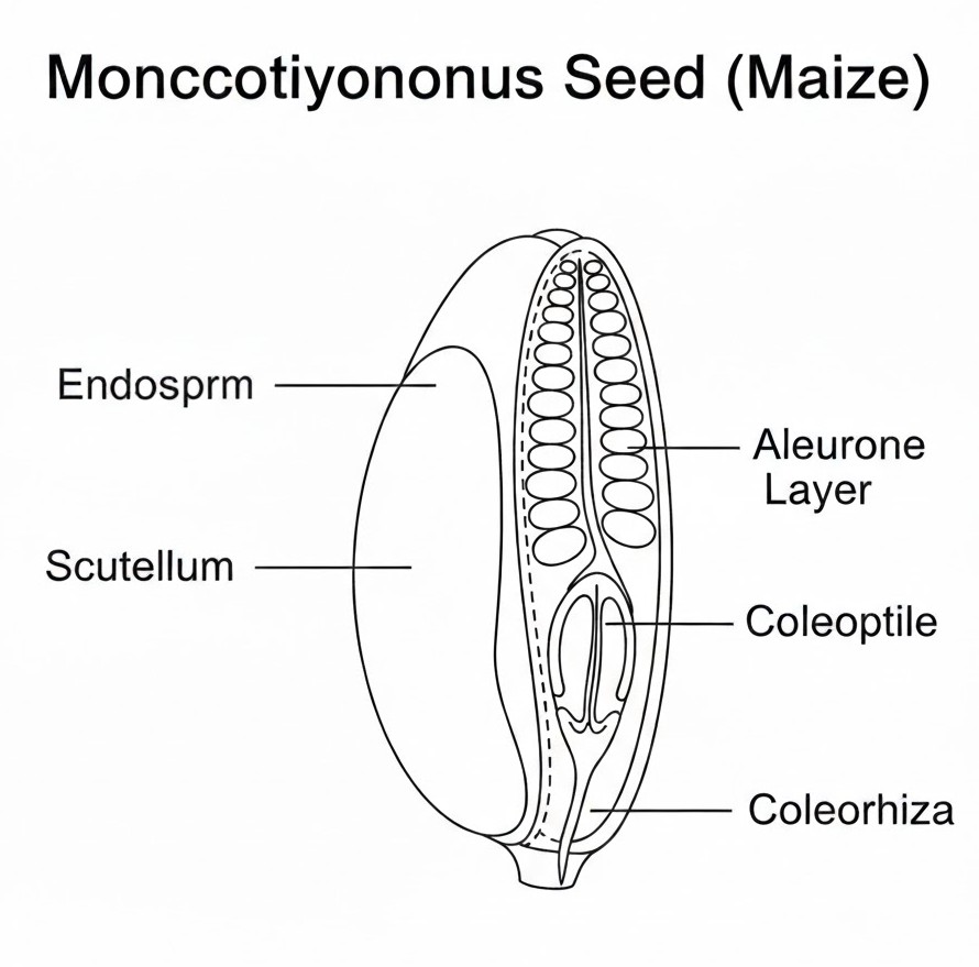
Monocots like maize usually have one shield-shaped cotyledon called a scutellum. They are mostly endospermous and have a special protein layer called the aleurone layer.
NEET Checkpoint:
The protective sheath of the radicle in monocot seeds is:
(a) Coleoptile
(b) Scutellum
(c) Coleorhiza
(d) Testa
✅ Answer: (c) Coleorhiza — Remember: Radicle has Rhiza.
Figure below:
A clear, labeled diagram of a monocotyledonous seed (maize) showing the endosperm, aleurone layer, scutellum, coleoptile, and coleorhiza.
Practice Questions:
1. In a maize seed, the plumule is enclosed within:
(a) Coleorhiza
(b) Coleoptile
(c) Scutellum
(d) Tegmen
✅ Answer: (b) Coleoptile. The plumule gets the "tile" while the radicle gets the "rhiza."
2. Which of the following is a non-endospermous dicot seed?
(a) Castor
(b) Maize
(c) Pea
(d) Coconut
✅ Answer: (c) Pea. Most dicots (except castor) use up their endosperm food during development.
Key Takeaway
- Fruits: Ripened ovaries; Drupes (mango/coconut) have one seed and a differentiated pericarp.
- Dicot Seeds: Two cotyledons; usually non-endospermous (e.g., pea, gram).
- Monocot Seeds: One cotyledon (scutellum); usually endospermous (e.g., maize).
12. Taxonomy of Flowering Plant Families
Listen up, junior! Ready to decode the "family drama" of plants? While your syllabus mentions several families, our main focus today—straight from the sources—is on the Brassicaceae and Solanaceae.
Brassicaceae (the Mustard family) is all about symmetry. Its floral formula is:
This tells us it's actinomorphic and bisexual. Imagine a plate of sarson da saag; that mustard plant is showing off a superior ovary and six stamens in two ranks ( $A{2+4}$).
NEET Checkpoint:
What does the symbol $\oplus$ represent?
(a) Normal fertilization
(b) Actinomorphic
(c) Pollination
(d) Double fertilization
✅ Answer: (b) Actinomorphic — It means the flower has radial symmetry, like a cricket ball!.
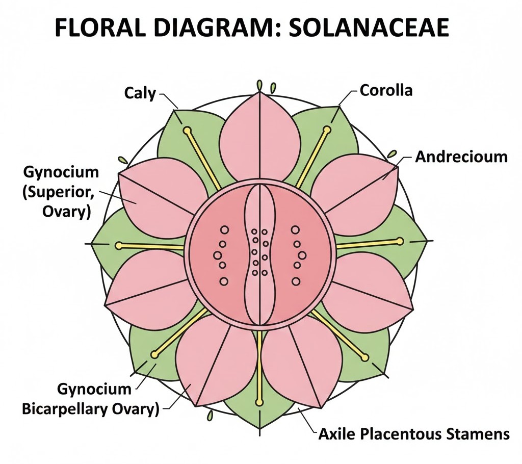
Next is the Solanaceae or "Potato family." These are the real MVPs of your kitchen—potatoes, tomatoes, and chillies. They have epipetalous stamens, meaning the stamens are attached to the petals like a pillion rider on a busy rickshaw. Their formula is:
They feature a persistent calyx (the green "cap" on a brinjal) and axile placentation.
NEET Checkpoint:
Which family is a source of medicine like Ashwagandha?
(a) Fabaceae
(b) Solanaceae
(c) Lamiaceae
(d) Asteraceae
✅ Answer: (b) Solanaceae — It's not just for aloo parathas; it has great medicinal value too!.
Figure below:
A clear floral diagram of Solanaceae showing five united sepals, five united petals with five epipetalous stamens, and a bicarpellary syncarpous superior ovary with axile placentation.
NEET Checkpoint:
In Solanaceae, the ovary is?
(a) Superior
(b) Inferior
(c) Half-inferior
(d) Epigynous
✅ Answer: (a) Superior — Indicated by the line below the $G$ ($\underline{G}$) in the formula.
Note: While Malvaceae, Leguminosae, Compositae, and Gramineae are mentioned in the unit overview, they aren't detailed in these specific pages.
Practice Questions:
1. The floral formula of Mustard belongs to which family?
(a) Solanaceae
(b) Brassicaceae
(c) Liliaceae
(d) Fabaceae
✅ Answer: (b) Brassicaceae — Remember the $K_{2+2}$ and $C_4$ arrangement!.
2. Which of the following is a characteristic of Solanaceae?
(a) Fibrous roots
(b) Epipetalous stamens
(c) Inferior ovary
(d) Parallel venation
✅ Answer: (b) Epipetalous stamens — The stamens are fused with the petals.
Key Takeaway
- Brassicaceae: $\oplus \underline{Q}^{7}K_{2+2} C_{4} A_{2+4}\underline{G}_{(2)}$ (e.g., Mustard).
- Solanaceae: $\oplus \underline{Q}^{7}K_{(5)}\widehat{C_{(5)}A_{5}}\underline{G}_{(2)}$; includes potato, tomato, and chilli.
13. Animal Tissues and Frog Morphology
Listen up, junior! Before we jump into the anatomy of our favorite pond-hopper, remember that all complex animals—including us—are built from just four basic tissue types: epithelial, connective, muscular, and neural. Think of these like the four main subjects for NEET; you need all of them to make the system work! These tissues organize into organs, which then team up to form organ systems, showing a perfect division of labour.
Now, let's talk about the star of the show: Rana tigrina, the common Indian bullfrog. These guys are poikilotherms (cold-blooded), meaning their body temperature fluctuates like your mood during a mock test. To survive the extreme Indian weather, they perform "summer sleep" (aestivation) and "winter sleep" (hibernation) in deep burrows. Talk about a long nap, yaar!
NEET Checkpoint:
Which of the following is a protective coloration used by frogs to hide?
(a) Hibernation
(b) Aestivation
(c) Mimicry
(d) Digestion
✅ Answer: (c) Mimicry — This is the camouflage that helps them blend into grasses to dodge enemies!
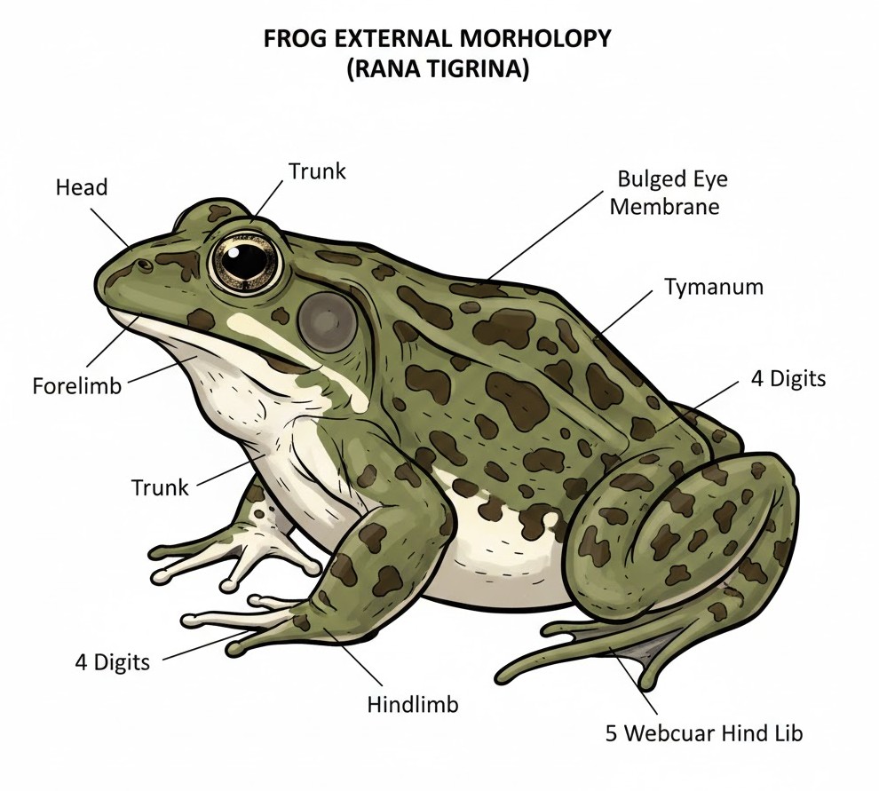
Morphology-wise, the frog is pretty sleek. Their skin is smooth and slippery due to mucus, and they actually absorb water through their skin instead of drinking it. The body is divided into just a head and trunk—notice that a neck and tail are completely absent. Their eyes are bulged and protected by a nictitating membrane while underwater.
Figure below:
A clear diagram of a frog (Rana tigrina) showing the external morphology: head, trunk, bulged eyes with nictitating membrane, tympanum, forelimbs with 4 digits, and larger muscular hind limbs with 5 webbed digits.
NEET Checkpoint:
How many digits are found on the forelimbs and hind limbs of a frog respectively?
(a) 5, 4
(b) 4, 5
(c) 4, 4
(d) 5, 5
✅ Answer: (b) 4, 5 — The hind limbs are larger and muscular, ending in five webbed digits to help with swimming!
NEET Checkpoint:
Which structure in the frog is responsible for receiving sound signals?
(a) Nictitating membrane
(b) Vocal sacs
(c) Tympanum
(d) Copulatory pad
✅ Answer: (c) Tympanum — This is the membranous ear seen on either side of the eyes.
Finally, keep an eye out for sexual dimorphism. Male frogs are the loud ones because they possess vocal sacs; they also have a copulatory pad on the first digit of their forelimbs. These features are absent in females, so now you know who's making all that noise in the rain!
Practice Question:
A frog's skin is always maintained in a moist condition. This is important because:
(a) It helps them drink water
(b) It is necessary for respiration
(c) It changes their colour
(d) It helps them walk
✅ Answer: (b) It is necessary for respiration — Frogs use their moist skin for cutaneous respiration, especially in water.
Key Takeaway
- Four Tissues: Epithelial, Connective, Muscular, Neural.
- Frog Species: Rana tigrina.
- Body: Head and Trunk only; Skin is moist/mucus-covered.
- Male Features: Vocal sacs and copulatory pads.
14. Frog Anatomy: Digestive, Respiratory, and Circulatory Systems
Alright, junior, let’s get into the "inner workings" of our favorite amphibian, Rana tigrina. Think of this as opening the bonnet of a car to see how the engine runs!
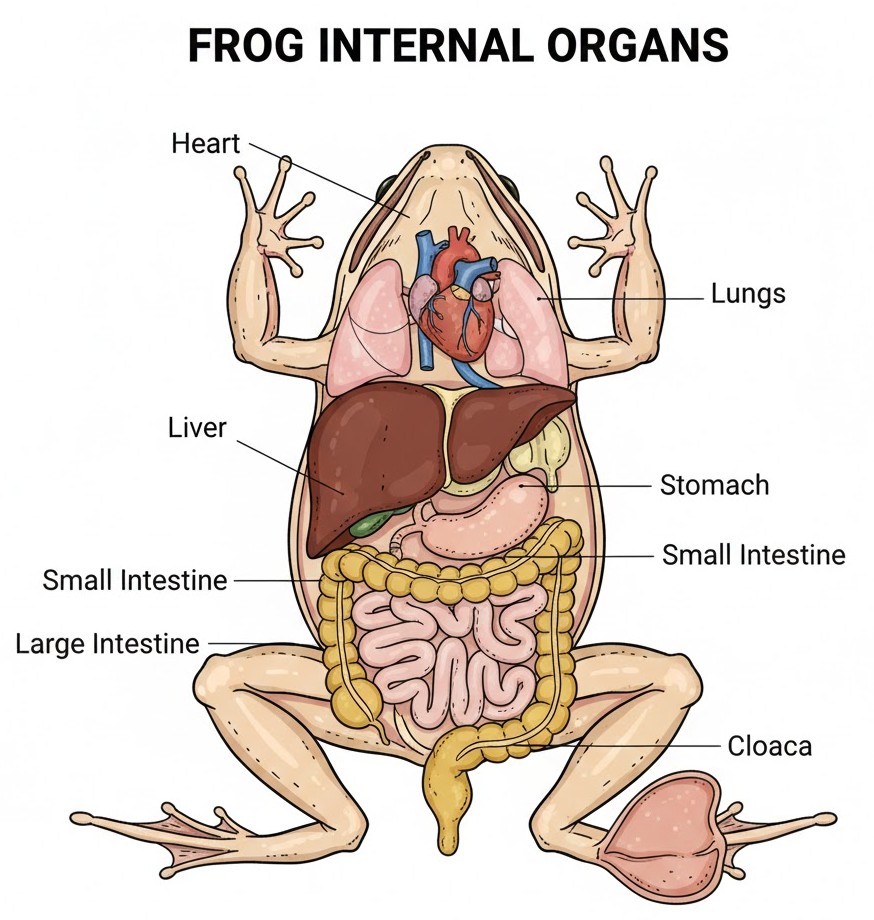
First up, the Digestive System. Because frogs are carnivores, they don't need a long, winding "highway" for digestion; their alimentary canal is actually quite short. Imagine a "fast-track" lane at a Delhi toll booth—quick and efficient! Their bilobed tongue snaps up prey, which then travels through the oesophagus to the stomach. Here, $HCl$ and gastric juices break it down into a semi-digested paste called chyme.
NEET Checkpoint:
Where does the final digestion of food take place in a frog?
(a) Stomach
(b) Small intestine
(c) Large intestine
(d) Buccal cavity
✅ Answer: (b) Small intestine — This is where the final breakdown happens before absorption via villi.
Next, let's talk about Respiration. Frogs are the ultimate multitaskers. In water, they use cutaneous respiration (breathing through skin), but on land, they can switch to their buccal cavity or pink, sac-like lungs (pulmonary respiration). During "summer sleep" (aestivation), they rely solely on their skin. It’s like having a smartphone that charges via cable, wireless, and solar power!
NEET Checkpoint:
Which respiratory method is used during hibernation?
(a) Pulmonary respiration
(b) Cutaneous respiration
(c) Branchial respiration
(d) Tracheal respiration
✅ Answer: (b) Cutaneous respiration — The skin is the only worker during those long winter naps.
Finally, the Circulatory System. Frogs have a closed system with a three-chambered heart (2 atria and 1 ventricle). They also have unique "VIP lanes" called portal systems: the hepatic portal system (between liver and intestine) and the renal portal system (between kidney and lower body). Interestingly, their RBCs are nucleated—so they’ve got a "brain" in every red cell!
Figure below:
A clear diagram showing the internal organs of a frog, specifically highlighting the heart, lungs, liver, and the short, looped alimentary canal ending in the cloaca.
NEET Checkpoint:
How many chambers are in a frog's heart?
(a) 2
(b) 3
(c) 4
(d) 5
✅ Answer: (b) 3 — Two atria to receive blood and one muscular ventricle to pump it out.
Practice Questions:
1. The hepatic portal system connects which two organs?
(a) Kidney and Heart
(b) Liver and Intestine
(c) Liver and Kidney
(d) Heart and Lungs
✅ Answer: (b) — Arre, it's a special connection just for the liver and gut!
2. Which of the following is TRUE for frog RBCs?
(a) Non-nucleated
(b) Contain haemoglobin and are nucleated
(c) Lack red pigment
(d) Are only found in the lymph
✅ Answer: (b) — Unlike us humans, these guys keep the nucleus in their RBCs!
Key Takeaway
- Digestive: Short canal (carnivore), $HCl$ in stomach, absorption in intestine.
- Respiratory: Skin (water/land/sleep) and Lungs (land).
- Circulatory: 3-chambered heart, closed system, presence of hepatic and renal portal systems.
15. Frog Anatomy: Nervous, Excretory, and Reproductive Systems
Arre junior, don't stress—understanding the frog’s "command centre" and "plumbing" is easier than it looks! The nervous system is highly evolved, featuring a Central Nervous System (CNS) (brain and spinal cord), a Peripheral Nervous System (PNS) (cranial and spinal nerves), and an Autonomic Nervous System (ANS). Think of the frog's brain like a busy pani puri stall—the fore-brain handles the orders (smell and logic), the mid-brain watches the crowd with its optic lobes, and the hind-brain (cerebellum and medulla) keeps the chef's balance!
NEET Checkpoint:
How many pairs of cranial nerves arise from a frog's brain?
(a) 8 pairs
(b) 9 pairs
(c) 10 pairs
(d) 12 pairs
✅ Answer: (c) $10 \text{ pairs}$ — These nerves help the brain communicate with various parts of the head.
Next up is the excretory system. Frogs have a pair of dark red, bean-like kidneys tucked away posteriorly. Inside, nephrons do the heavy lifting of filtering blood. Because they excrete urea, frogs are ureotelic. It’s like a Mumbai local train—everything, from nitrogenous waste to reproductive cells, eventually gathers at the cloaca 'station' before heading out to the world!
NEET Checkpoint:
Why is the frog described as "ureotelic"?
(a) It excretes uric acid
(b) It excretes ammonia
(c) It excretes urea
(d) It excretes both urea and uric acid
✅ Answer: (c) Because it excretes urea — Nitrogenous wastes are carried by blood to the kidneys for separation.
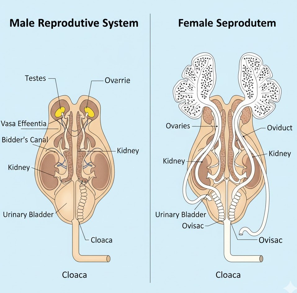
Now for the reproductive system. Males have yellowish testes that send sperms through $10 \text{--} 12$ vasa efferentia into Bidder's canal. In females, ovaries produce $2500 \text{--} 3000$ ova at a time, which pass through separate oviducts. Fertilisation is external, happening in water—essentially a massive aquatic 'pool party' where eggs become tadpoles.
NEET Checkpoint:
What is the common passage for urine and sperms in male frogs?
(a) Ureter
(b) Urinogenital duct
(c) Cloaca
(d) Urinary bladder
✅ Answer: (b) Urinogenital duct — This duct emerges from the kidney and opens into the cloaca.
Figure below:
A clear, side-by-side anatomical diagram of male and female frog reproductive systems. The male side should show testes, vasa efferentia, and Bidder's canal within the kidney. The female side should show ovaries and separate oviducts. Both systems should clearly terminate at the cloaca.
Practice Question:
In a male frog, the vasa efferentia enter the kidneys and open into:
(a) Ureter
(b) Bidder's canal
(c) Urinary bladder
(d) Oviduct
✅ Answer: (b) Bidder's canal — This is a specific structure within the male kidney that communicates with the urinogenital duct.
Key Takeaway:
- Nervous System: Comprises CNS, PNS ($10 \text{ pairs}$ of cranial nerves), and ANS.
- Excretory System: Pair of kidneys; frogs are ureotelic (excrete urea).
- Reproductive System: Males have a urinogenital duct; females lay thousands of eggs; fertilisation is external in water.