1. Introduction to Human Physiological Systems
All our organ systems need to chat and work together perfectly to maintain homeostasis. Think of homeostasis as keeping the body's internal environment super steady, like maintaining the AC temperature perfectly during a Delhi heatwave. Coordination means two or more organs interact and complement each other's functions.
This synchronisation act is jointly managed by two powerhouse systems: the Neural System and the Endocrine System. Notice this: the neural system provides rapid, point-to-point coordination. It’s like sending a WhatsApp message—instant! The endocrine system, however, uses non-nutrient chemicals called hormones for chemical integration. That’s like sending a post parcel—slower, but the effect spreads wider, regulating cellular functions continuously.
NEET Checkpoint:
Which systems jointly coordinate and integrate physiological activities?
(a) Skeletal and Muscular
(b) Neural and Endocrine
(c) Digestive and Excretory
(d) Respiratory and Circulatory
✅ Answer: (b) Neural and Endocrine — Bada funda: they work together to synchronise everything and maintain homeostasis.
Imagine a bike messenger (neural system) delivering a rush document instantly. That's quick, point-to-point action! Now, think about the gradual, body-wide changes during puberty; that’s managed by the slower, systemic action of the endocrine system using hormones.
This entire unit (Human Physiology) dives deep into these life processes, describing things like gas exchange (Chapter 14), blood circulation (Chapter 15), and locomotion (Chapter 17) in cellular and molecular terms. The final chapters point to the organismic-level regulation provided by the neural and endocrine systems. Speaking of gas exchange, the first system we tackle is respiration, where we exchange atmospheric $O_2$ with $CO_2$ produced by our cells—that’s breathing.
NEET Checkpoint:
Hormones are best described as:
(a) Cellular structures
(b) Enzymes
(c) Intercellular messengers
(d) Structural proteins
✅ Answer: (c) Intercellular messengers — They are non-nutrient chemicals produced in trace amounts that act as messengers between cells.
Practice Questions
- If you suddenly step on a rickshaw brake, the rapid signal transmission to your leg muscles is primarily handled by the:
(a) Endocrine system
(b) Excretory system
(c) Neural system
(d) Cardiac system
✅ Explanation: (C) The neural system is specialized for quick, electrical signalling providing rapid, point-to-point coordination.
2. The function of regulating body temperature and urges for eating and drinking is associated with which part of the forebrain?
(a) Cerebrum
(b) Thalamus
(c) Pons
(d) Hypothalamus
✅ Explanation: (d) The hypothalamus contains centres which control body temperature, and the urge for eating and drinking.
Key Takeaway
Homeostasis keeps the body stable by relying on the synchronization of all organ activities. This essential job is performed jointly by the rapid, point-to-point Neural System and the slower, chemical Endocrine System.
2. Structure and Mechanism of Human Breathing
We need to understand the super-efficient highway our body uses for gas exchange. The human respiratory system is basically a complex pipeline starting with the external nostrils, which lead into the nasal chamber via the nasal passage. This path continues through the pharynx, which is interesting because it’s a common passage for both food and air. From there, we pass through the cartilaginous larynx—our natural sound box!—before hitting the main windpipe, the trachea.
The trachea is a straight tube that extends down and divides into the right and left primary bronchi. These bronchi undergo repeated divisions to form secondary and tertiary bronchi and bronchioles, finally ending up in very thin terminal bronchioles. Eventually, these terminal bronchioles give rise to numerous very thin, irregular-walled, and highly vascularised bag-like structures called alveoli. This entire branching network comprises the lungs.
Notice this: The path from the external nostrils up to the terminal bronchioles is the conducting part. Its job is crucial: transport the atmospheric air to the alveoli, clear foreign particles (no kachra allowed!), humidify it, and warm the air to body temperature. The alveoli and their ducts are the superstars—they form the exchange part, which is the actual site of $\text{O}_2$ and $\text{CO}_2$ diffusion between the blood and the atmospheric air.
NEET Checkpoint:
The conducting part of the human respiratory system clears the air of foreign particles and performs which two other functions?
(a) Cooling and filtering
(b) Humidifying and warming
(c) Gas exchange and transport
(d) Sound production and transport
✅ Answer: (b) Humidifying and warming — The conducting part transports air, clears foreign particles, humidifies, and also brings the air to body temperature.
The mechanism of breathing (pulmonary ventilation) involves drawing in atmospheric air (inspiration) and releasing $\text{CO}_2$-rich alveolar air (expiration). It all depends on creating a pressure gradient. Think of it like sucking pani puri water with a straw: you decrease the pressure in your mouth below the outside pressure!
For inspiration, the pressure within the lungs (intra-pulmonary pressure) must be less than the atmospheric pressure—a negative pressure situation. Inspiration is initiated by the contraction of the diaphragm, increasing the thoracic chamber volume in the antero-posterior axis. Simultaneously, the contraction of the external inter-costal muscles lifts the ribs and sternum, increasing the volume in the dorso-ventral axis. This overall volume increase leads to an increase in pulmonary volume, which drops the intra-pulmonary pressure, forcing air inwards. Bas ho gaya!
Figure below:
A clear diagram showing the human respiratory pathway from the larynx to the alveoli, and a separate diagram illustrating the mechanism of breathing with the diaphragm contracted (inspiration) and relaxed (expiration).
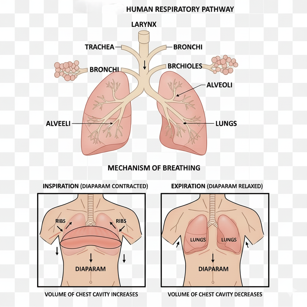
For expiration, things relax. The diaphragm and the inter-costal muscles relax, returning the chest structures to their normal positions. This relaxation reduces the thoracic and pulmonary volume, which increases the intra-pulmonary pressure to slightly above the atmospheric pressure, causing the air to be expelled.
NEET Checkpoint:
Inspiration is considered an active process primarily due to:
(a) Passive elastic recoil of the lungs
(b) Contraction of the diaphragm and external intercostal muscles
(c) Relaxation of abdominal muscles
(d) High alveolar $\text{pCO}_2$
✅ Answer: (b) Contraction of the diaphragm and external intercostal muscles — Inspiration is initiated by the contraction of these specialized muscles.
Practice Question:
If the intra-pulmonary pressure is represented as $P_{\text{lung}}$ and the atmospheric pressure is $P_{\text{atm}}$, what condition correctly describes normal inspiration?
(a) $P_{\text{lung}} > P_{\text{atm}}$
(b) $P_{\text{lung}} < P_{\text{atm}}$
(c) $P_{\text{lung}} = P_{\text{atm}}$
(d) $P_{\text{lung}} = 40 \text{mm Hg}$
✅ Answer: (b) $P_{\text{lung}} < P_{\text{atm}}$ — Inspiration occurs if the pressure within the lungs is less than the atmospheric pressure, creating a negative gradient.
Key Takeaway
Breathing is driven entirely by creating pressure gradients. Inspiration is an active process initiated by the contraction of the diaphragm and external intercostal muscles, increasing thoracic volume and creating negative pressure. Expiration normally involves the relaxation of these muscles, decreasing volume and increasing pressure to expel $\text{CO}_2$-rich air. The conducting pathway cleans and conditions the air before it reaches the alveoli, the site of diffusion.
3. Respiratory Volumes, Gas Transport, and Respiratory Disorders: The Oxygen Hustle
Hey juniors! Ready to clear up the funda behind how much air we push around and how our blood pulls off the gas exchange magic? Don't stress, this section is pure gold for NEET!
We start with volumes. The air you breathe normally is the Tidal Volume ($\mathbf{TV}$), about $500~mL$. If you force yourself to inhale, that extra air is Inspiratory Reserve Volume ($\mathbf{IRV}$) ($2500$ to $3000~mL$). Similarly, the extra air you can force out is Expiratory Reserve Volume ($\mathbf{ERV}$) ($1000$ to $1100~mL$). Even after that forceful exhale, some air stays back—that's the Residual Volume ($\mathbf{RV}$) ($1100$ to $1200~mL$). Combining these gives us Pulmonary Capacities (like Vital Capacity or Total Lung Capacity), essential for clinical diagnosis.
NEET Checkpoint:
Which capacity includes Tidal Volume, IRV, and ERV?
(a) Total Lung Capacity
(b) Functional Residual Capacity
(c) Vital Capacity
(d) Inspiratory Capacity
✅ Answer: (c) Vital Capacity — This volume represents the maximum air that can be breathed in after a forced expiration ($VC = ERV + TV + IRV$).
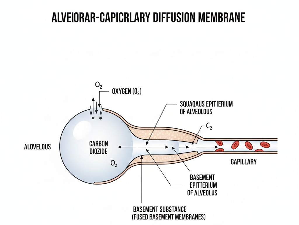
The actual gas exchange is straight-up simple diffusion. Gases move across the alveolar membrane primarily based on the partial pressure gradient. For example, $O_{2}$ moves from the high pressure in the alveoli ($p{O_{2}}=104~mm~Hg$) to the lower pressure in deoxygenated blood ($p{O_{2}}=40~mm~Hg$). Notice this—the diffusion membrane is super thin, less than a millimetre thick, comprising three layers: alveolar squamous epithelium, capillary endothelium, and the basement substance.
Figure below:
A clear diagram showing the alveolar-capillary diffusion membrane, highlighting the three layers: squamous epithelium of the alveoli, endothelium of the capillary, and the basement substance between them.
For transport, $97\%$ of $O_{2}$ hitches a ride on haemoglobin (Hb) in RBCs, forming oxyhaemoglobin. This binding is favoured in the alveoli due to high $\mathbf{p{O_{2}}}$ and low $\mathbf{p{CO_{2}}}$. Carbon dioxide ($CO_{2}$) is mostly transported as bicarbonate ($\mathbf{HCO_{3}^{-}}$, about $70\%$). It needs the superstar enzyme carbonic anhydrase (very high concentration in RBCs) to facilitate the reaction:
The rest of $CO_{2}$ binds to Hb as carbamino-haemoglobin ($20-25\%$).
NEET Checkpoint:
$CO_{2}$ is primarily transported in the blood as:
(a) Carbamino-haemoglobin
(b) Dissolved in plasma
(c) Bicarbonate ions
(d) Dissolved in RBCs
✅ Answer: (c) Bicarbonate ions — Approximately $70\%$ of $CO_{2}$ is carried this way.
Now, the control system! Our breathing rhythm is primarily managed by the respiratory rhythm centre in the medulla. Fun fact: This centre is way more sensitive to changes in $\mathbf{CO_{2}}$ and $\mathbf{H^{+}}$ concentration than it is to $O_{2}$. $O_{2}$'s role here? Insignificant, yaar!
Imagine you’re waiting in line for pani puri (high $CO_{2}$ concentration, like in tissues). The longer the line, the more stressed (activated) you get, forcing the system (medulla) to signal faster elimination.
Finally, the respiratory roadblocks! Asthma involves difficulty breathing (wheezing) because of inflammation in the bronchi/bronchioles. Emphysema is a nasty chronic disorder where the alveolar walls get damaged, cutting down the respiratory surface—often linked to smoking. Don't forget Occupational Respiratory Disorders, which cause fibrosis (proliferation of fibrous tissues) due to prolonged dust exposure. Always mask up if you work there!
NEET Checkpoint:
Emphysema is characterized by:
(a) Inflammation of bronchioles
(b) Damage to alveolar walls
(c) Accumulation of uric acid
(d) Bone degeneration
✅ Answer: (b) Damage to alveolar walls — This leads to a decreased respiratory surface.
Practice Question:
If $100~mL$ of oxygenated blood typically delivers about $5~mL$ of $O_{2}$ to the tissues, how many $mL$ of $CO_{2}$ does $100~mL$ of deoxygenated blood deliver to the alveoli?
(a) $3~mL$
(b) $4~mL$
(c) $5~mL$
(d) $7~mL$
✅ Explanation: $100~mL$ of deoxygenated blood delivers approximately $4~mL$ of $CO_{2}$ to the alveoli.
Key Takeaway
Volumes like $\mathbf{TV}$ ($500~mL$) define normal breathing. Gas movement relies purely on partial pressure gradients. Oxygen transport ($97\%$ via $\mathbf{Hb}$) is favoured by high $\mathbf{pO_{2}}$. $\mathbf{CO_{2}}$ transport ($70\%$ as $\mathbf{HCO_{3}^{-}}$) uses carbonic anhydrase. The Medulla is the boss, tuning respiration based mainly on $\mathbf{CO_{2}}$ and $\mathbf{H^{+}}$ levels. Disorders include Asthma (inflammation), Emphysema (alveolar damage), and Occupational fibrosis.
4. Composition of Blood and Blood Grouping
It's classified as a special connective tissue that functions as the body’s main transport system. It's essentially split into two main components: the fluid matrix called plasma, which makes up nearly $55\%$ of the volume, and the solid components called formed elements, which constitute the remaining $45\%$.
Plasma is a viscous, straw-coloured fluid, mostly $(90-92\%)$ water. The key proteins in plasma are super important: fibrinogen is mandatory for blood clotting; globulins are our defense squad, playing a major role in immune mechanisms; and albumins are the paaniwallah managing the water balance (osmotic balance). Don't forget that if you strip away the clotting factors from plasma, what remains is called serum. Now, that’s a quick funda cleared!
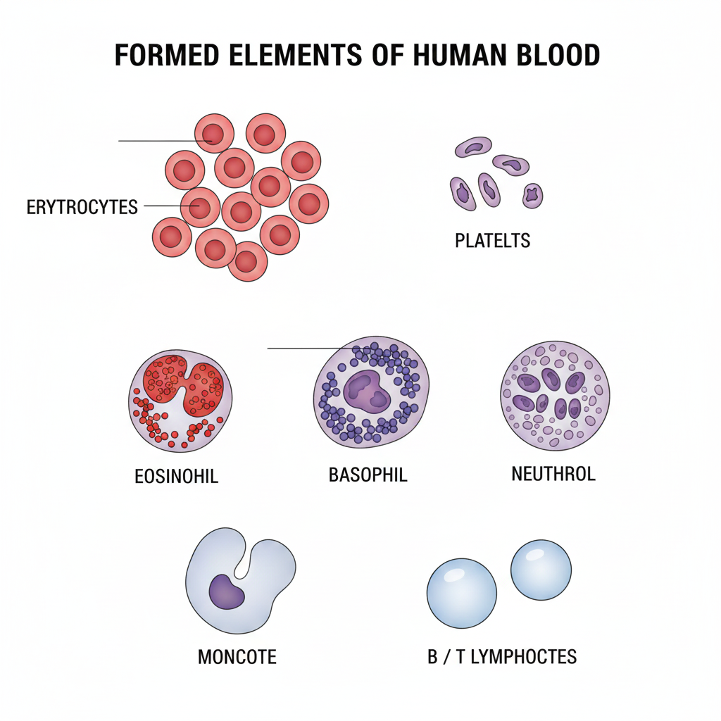
The formed elements include Erythrocytes (RBCs), Leucocytes (WBCs), and Platelets. RBCs are the gas transport experts because they contain the iron-containing pigment haemoglobin. They live for about $120$ days before being destroyed in the spleen, which is famously called the graveyard of RBCs.
WBCs (Leucocytes) are our nucleated defense fighters. They are split into granulocytes (Neutrophils, Eosinophils, Basophils) and agranulocytes (Lymphocytes, Monocytes). Notice this: Neutrophils $(60-65\%)$ and Monocytes $(6-8\%)$ are the phagocytic cells, ready to swallow foreign organisms. Basophils are the least common $(0.5-1\%)$, but they secrete substances like histamine necessary for inflammatory reactions. Platelets, which are cell fragments from megakaryocytes, are crucial for starting the coagulation process.
Figure below:
A clear diagram showing the formed elements in blood, including erythrocytes, platelets, eosinophils, basophils, neutrophils, monocytes, and B/T lymphocytes.
Imagine your study group preparing for NEET: RBCs are the textbooks carrying knowledge $(O_{2})$, WBCs are your friends defending against distractions (defense), and platelets are the highlighters stopping information leak (clotting) when things get tough!
NEET Checkpoint:
Which category of formed elements includes cells responsible for immune responses?
(A) Erythrocytes
(B) Thrombocytes
(C) Granulocytes
(D) Lymphocytes
✅ Answer: (D) Lymphocytes — These are a type of agranulocyte WBC responsible for the immune responses of the body.
Blood Grouping Funda
The two main systems for blood safety are ABO and Rh. ABO grouping is determined by the presence or absence of A and B antigens on the RBC surfaces. The trick here: individuals with Group AB blood have both A and B antigens but no antibodies, making them universal recipients. Conversely, Group O lacks both antigens but has anti-A and anti-B antibodies, making them the universal donors.
The Rh grouping involves the presence ($Rh^{+ve}$) or absence ($Rh^{-ve}$) of the Rh antigen. This is super critical for pregnancy! If an $Rh^{-ve}$ mother carries an $Rh^{+ve}$ foetus, her body might start preparing antibodies during the first delivery. In subsequent pregnancies, these maternal $\text{Rh}$ antibodies can leak into the foetal blood and destroy its RBCs, causing the severe condition called erythroblastosis foetalis.
NEET Checkpoint:
Which component of plasma helps maintain the osmotic balance?
(A) Albumins
(B) Fibrinogen
(C) Electrolytes
(D) Globulins
✅ Answer: (A) Albumins — These proteins specifically help in maintaining the osmotic balance of the blood.
NEET Checkpoint:
The process of blood coagulation is initiated by:
(A) Red blood cells (RBCs)
(B) White blood cells (WBCs)
(C) Plasma proteins
(D) Platelets (thrombocytes)
✅ Answer: (D) Platelets (thrombocytes) — They release a variety of substances involved in the cascade of clotting mechanisms.
Practice Question:
Which blood group is considered the universal recipient and why?
(a) Group A, because it lacks A antigens.
(b) Group O, because it lacks both A and B antigens.
(c) Group AB, because its plasma lacks anti-A and anti-B antibodies.
(d) Group B, because it lacks anti-A antibodies.
✅ Answer: (c) Group AB, because its plasma lacks anti-A and anti-B antibodies — Since it has no antibodies against A or B, it can accept blood from any group without clumping.
Key Takeaway
Blood is special connective tissue $(55\%\ \text{Plasma} + 45\%\ \text{Formed Elements})$. Plasma proteins like Fibrinogen (clotting) and Albumins (osmotic balance) are key. RBCs carry $\text{Hb}$ and are destroyed in the spleen. WBCs (like phagocytic Neutrophils/Monocytes) handle defense. ABO grouping relies on antigens/antibodies, making 'O' the universal donor and 'AB' the universal recipient. Rh incompatibility between $Rh^{-ve}$ mother and $Rh^{+ve}$ foetus leads to erythroblastosis foetalis.
5. Coagulation of Blood and Lymphatic System
Let's tackle the funda of how your body prevents a tiny scratch from becoming a major issue: Blood Coagulation. When you get a trauma or injury (like scraping your knee while falling off your scooter), your blood quickly undergoes coagulation or clotting to prevent excessive loss of blood from the body. Don't stress, yaar—it’s an amazing defense mechanism!
The resulting dark reddish-brown scum we call a clot is primarily a network of threads called fibrins. These threads trap the dead and damaged formed elements of the blood. Now, how are fibrins formed? It’s a classic activation cascade! Fibrins come from inactive fibrinogen (which is a major protein found in the plasma) using the enzyme thrombin.
NEET Checkpoint:
Plasma without clotting factors is specifically called:
(a) Lymph
(b) Serum
(c) Tissue Fluid
(d) Buffy Coat.
✅ Answer: (b) Serum — Remember, plasma without clotting factors (like fibrinogen) is specifically defined as serum.
And here’s the trick: thrombin itself starts as inactive prothrombin in the plasma. A complex enzyme called thrombokinase is required to facilitate this conversion. This enzyme complex is formed by a series of linked enzymic reactions (a cascade process) involving a number of inactive factors in the plasma. Factors released by both platelets and injured tissues can initiate this mechanism. Notice this, yaar: Calcium ions ($Ca^{++}$) play a very important role in this whole clotting mechanism. Without proper$Ca^{++}$ levels, your blood clotting funda goes haywire!
Now let's quickly shift gears to Lymph (Tissue Fluid). This fluid is super important for exchange between blood and cells. Imagine a pani puri stall (capillaries) where water along with many small water-soluble substances (like nutrients and gases) move out into the crowded tissue spaces. This leaked fluid is the interstitial fluid or tissue fluid. This fluid is similar to plasma in its mineral distribution but leaves the larger proteins and most formed elements inside the capillaries.
An elaborate network, the lymphatic system, collects this tissue fluid and drains it back into the major veins. The fluid inside the lymphatic system is called lymph. Lymph is a colourless fluid that contains specialized lymphocytes. And that’s cool, right? These specialized cells are primarily responsible for the body's immune responses. Lymph also acts as an important carrier for nutrients and hormones, notably facilitating the absorption of fats through the lacteals present in the intestinal villi.
NEET Checkpoint:
Which substance is specifically absorbed via lymph in intestinal villi?
(a) Glucose
(b) Water
(c) Fats
(d) Amino Acids.
✅ Answer: (c) Fats — Lymph is an important carrier, especially for fats absorbed through lacteals in the intestinal villi.
Practice Question:
The cascade process of blood coagulation is initiated by factors released by:
(a) Only tissues at the injury site
(b) Only platelets
(c) Both tissues and platelets at the injury site
(d) Albumins in the plasma.
✅ Answer: (c) Both tissues and platelets at the injury site — An injury stimulates platelets to release factors, and tissues at the site of injury also release factors, both initiating coagulation.
NEET Checkpoint:
The ultimate function of the specialized lymphocytes found in lymph is:
(a) Osmotic balance
(b) Transporting oxygen
(c) Immune responses
(d) Gluconeogenesis.
✅ Answer: (c) Immune responses — Lymph contains specialised lymphocytes that are responsible for the body’s immune responses.
Key Takeaway
Clotting is a $Ca^{++}$ dependent cascade where inactive prothrombin converts to thrombin via thrombokinase, which then converts fibrinogen into the clot-forming fibrin. Lymph, the tissue fluid, handles drainage and houses key lymphocytes for immunity.
6. The Heartbeat Hustle: Circulation, Cycles, and Cardiac Crisis
let's clear up the funda of how our internal pumping system works, because this topic is a NEET classic! Humans possess a closed circulatory system where blood is always circulated through a closed network of vessels, which is super efficient for regulation. Our heart is a muscular, 4-chambered organ, shielded by a double-walled pericardium. We have two smaller upper chambers, the atria, and two larger lower chambers, the ventricles.
The core funda of heart function is that it’s myogenic—it auto-regulates its activity using specialized nodal tissue. The boss of this tissue is the Sino-atrial Node (SAN), which is why it’s called the pacemaker. It sets the rhythm by generating action potentials at a rate of 70–75/min (average $72~beats~min^{-1}$).
The Cardiac Clockwork
The Cardiac Cycle is the sequential and cyclical event involving the contraction (systole) and relaxation (diastole) of the chambers. Notice this entire process wraps up in a quick $0.8~seconds$! During each cycle, each ventricle pumps out about $70~mL$ of blood, known as the stroke volume.
NEET Checkpoint:
What is the average duration of one complete cardiac cycle?
(a) $72~seconds$
(b) $0.5~seconds$
(c) $0.8~seconds$
(d) $5~litres$
✅ Answer: (c) $0.8~seconds$ — This is the duration required for one complete sequence of atrial and ventricular systole and diastole.
We calculate the Cardiac Output (CO), the volume of blood pumped per ventricle per minute, using this key formula:
This averages to about $5000~mL$ or $5~litres$ in a healthy individual.
Think of the Double Circulation system like a public bus network with two distinct lines. The Pulmonary circulation line runs strictly from the Right Ventricle to the Lungs to pick up oxygen, returning to the Left Atrium. The Systemic circulation is the main line, starting from the Left Ventricle and running to all body tissues before returning the deoxygenated blood to the Right Atrium.
Reading the Electrical Chart (ECG)
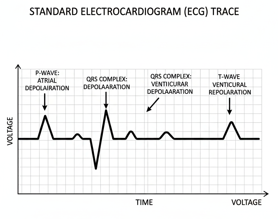
The ECG (Electrocardiogram) is a graphical representation that maps the heart’s electrical activity. Don’t mix up the waves, bro! The P-wave shows atrial depolarisation (contraction signal). The famous spike, the QRS complex, represents ventricular depolarisation. Lastly, the T-wave signifies ventricular repolarisation (relaxation).
Figure below:
A standard ECG trace showing the P-wave, QRS complex, and T-wave corresponding to atrial and ventricular depolarisation and repolarisation.
NEET Checkpoint:
Which ECG component represents ventricular repolarisation?
(a) P-wave
(b) QRS complex
(c) T-wave
(d) U-wave
✅ Answer: (c) T-wave — This wave shows the ventricles returning from the excited state back to normal.
The heart’s activity is regulated by a special neural centre in the medulla oblongata via the Autonomic Nervous System (ANS), and it can also be boosted by adrenal medullary hormones.
Cardiac Crisis (Disorders)
Be careful about these cardiac disorders, they are important:
- Hypertension: High blood pressure. If repeated checks show $140/90~mm~Hg$ or higher, that’s hypertension.
- Coronary Artery Disease (CAD): Narrowing of vessels supplying the heart muscle, typically due to deposits of calcium, fat, and cholesterol (atherosclerosis).
- Angina (Angina Pectoris): Acute chest pain occurring when the heart muscle isn't getting adequate oxygen.
- Heart Failure: The heart cannot pump blood effectively enough to meet the body's demands.
Practice Question:
CAD affects the vessels that supply blood directly to the heart muscle. The deposits that cause this disease include:
(a) Sodium, chloride, and water
(b) Calcium, fat, and cholesterol
(c) Urea and uric acid
(d) Plasma proteins
Answer: (b) Calcium, fat, and cholesterol. CAD is caused by deposits of calcium, fat, cholesterol, and fibrous tissues that narrow the artery lumen.
NEET Checkpoint:
The condition characterised by acute chest pain due to insufficient oxygen reaching the heart muscle is called:
(a) Heart Failure
(b) Hypertension
(c) Angina
(d) Arteriosclerosis
✅ Answer: (c) Angina — Angina is acute chest pain caused by inadequate oxygen reaching the heart muscle.
Key Takeaway
Cardiac Output is Stroke Volume ($\approx 70~mL$) $\times$ Heart Rate ($\approx 72/min$), averaging $5~litres$. The SAN is the pacemaker. ECG waves track electrical events: P (Atrial Depolarisation), QRS (Ventricular Depolarisation), T (Ventricular Repolarisation).
7. Modes of Excretion and Human Excretory System Structure
Let’s clear up the funda of how our body (and others!) gets rid of the trash, especially those pesky nitrogenous wastes. This topic is super important for understanding how we maintain water balance, so listen up!
Animals eliminate major nitrogenous wastes in three forms: ammonia, urea, and uric acid. Here’s the trick: ammonia is the most toxic form and demands a large amount of water for elimination. Because of this massive water requirement, animals that excrete ammonia are called ammonotelic. Examples include most bony fishes, aquatic amphibians, and aquatic insects.
NEET Checkpoint:
Which nitrogenous waste requires the minimum loss of water for elimination?
(a) Ammonia
(b) Urea
(c) Uric acid
(d) Amino acids
✅ Answer: (c) Uric acid — Least toxic, needs minimum water! Think of desert animals conserving every drop, yaar.
On the flip side, uric acid is the least toxic and can be removed with minimum water loss, often as a pellet or paste. Animals following this path are uricotelic, like reptiles, birds, and land snails. Now, for the middle path, we have ureotelism. Mammals (that's us!), many terrestrial amphibians, and marine fishes convert toxic ammonia into less toxic urea in their liver for excretion. This is why we are classified as ureotelic. It's like having a water filter system—ammonia needs the full flood (ammonotelism), while urea uses a measured splash (ureotelism).
Our own excretory system, the human urinary system, is straightforward. It consists of a pair of kidneys, one pair of ureters, a urinary bladder, and a urethra. These reddish-brown, bean-shaped kidneys have two distinct zones: an outer cortex and an inner medulla. Notice that the medulla isn’t a smooth layer; it's divided into conical masses called medullary pyramids that project into funnel-shaped spaces called calyces.
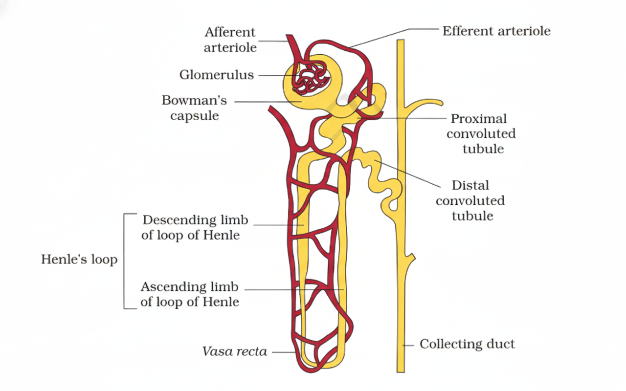
The real hero here is the nephron—the functional unit of the kidney. We have about one million of these complex tubular structures per kidney. Each nephron has two parts: the glomerulus and the renal tubule. The tubule starts with a double-walled, cup-like structure, the Bowman's capsule, which wraps around the glomerulus. Together, the glomerulus and the Bowman’s capsule form the Malpighian body (or renal corpuscle).
NEET Checkpoint:
The Malpighian body is formed by the combination of which two structures?
(a) PCT and DCT
(b) Glomerulus and Henle’s Loop
(c) Glomerulus and Bowman’s capsule
(d) Afferent and Efferent arteriole
✅ Answer: (c) Glomerulus and Bowman’s capsule — Easy peasy, just remember the twin structures.
The renal tubule then coils up dramatically to form the Proximal Convoluted Tubule (PCT), drops down into a hairpin-shaped Henle’s loop (HL), and finally coils again as the Distal Convoluted Tubule (DCT).
Figure below:
A clear diagram showing the overall structure of a nephron, detailing the Bowman's capsule, Glomerulus, PCT, Henle’s Loop (descending and ascending limbs), DCT, and the Collecting Duct.
NEET Checkpoint:
Which type of animals typically retain some urea in their kidney matrix to maintain osmolarity?
(a) Aquatic insects
(b) Bony fishes
(c) Mammals
(d) Birds
✅ Answer: (c) Mammals — This helps us maintain a desirable internal concentration, which is a key adaptation for terrestrial life.
Practice Questions:
1. The renal tubule segment that follows the Bowman's capsule is the:
(a) Henle's Loop
(b) Distal Convoluted Tubule
(c) Glomerulus
(d) Proximal Convoluted Tubule (PCT)
✅ Answer: (d) Proximal Convoluted Tubule (PCT) — The tubule starts at Bowman's capsule and continues directly into the highly coiled PCT.
2. In the human kidney, the medullary pyramids project into the:
(a) Cortex
(b) Hilum
(c) Calyces
(d) Urethra
✅ Answer: (c) Calyces — The medulla forms pyramids that project into the calyces.
Key Takeaway
The core 'funda' of excretion is dealing with nitrogenous waste, where toxicity (ammonia $>$ urea $>$ uric acid) dictates the water needed. Our functional unit is the nephron, composed of the Malpighian body (Glomerulus + Bowman's capsule) and the renal tubule (PCT, HL, DCT). Now that you know the structure, wait till you see how this complex machinery forms urine!
8. Urine Formation and Tubular Function
Making urine is a three-step mastery process inside the nephrons: glomerular filtration, reabsorption, and secretion.
First up is glomerular filtration, which we call ultra filtration because it’s so fine!. This process is powered by the glomerular capillary blood pressure. Blood is filtered across three layers: the endothelium of the glomerular blood vessels, the epithelium of Bowman's capsule (which has filtration slits or slit pores formed by podocytes), and the basement membrane connecting them. Almost everything in the plasma, except the proteins, gets filtered out into Bowman’s capsule.
Now, check out this amazing funda: the Glomerular Filtration Rate (GFR) in a healthy person is around $125~ml/minute$—that’s a massive $180$ litres per day!. Since we only excrete about $1.5$ litres of urine, this means a jaw-dropping $99$ per cent of that filtrate must be rescued via reabsorption by the renal tubules. Imagine if a cricket team had $180$ runs to chase but only let $1.5$ runs slip through—that's incredible efficiency!
NEET Checkpoint:
The GFR value of $180$ L/day means:
(a) The amount of urine excreted per day.
(b) The maximum blood pumped by the heart.
(c) The amount of filtrate formed per day.
(d) The water reabsorbed in the PCT.
✅ Answer: (c) The amount of filtrate formed per day — This is the total volume filtered, not the final urine volume.
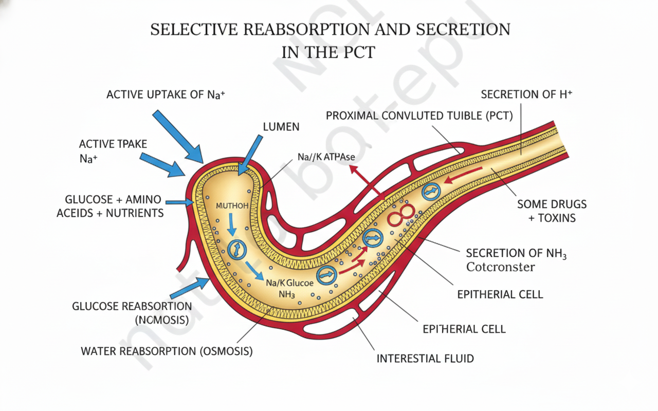
Reabsorption involves both active and passive mechanisms. Essential nutrients like glucose, amino acids, and $Na^{+}$ are actively reabsorbed. But it’s not just about taking things back; tubular secretion is the other vital mechanism! Tubular cells throw waste substances, specifically $H^{+}$ (hydrogen ions), $K^{+}$ (potassium ions), and ammonia ($NH_{3}$), directly into the filtrate. This secret weapon is necessary for keeping the ionic and acid-base balance ($pH$) of the body fluids steady.
The Proximal Convoluted Tubule (PCT) is like the chief warehouse, doing most of the heavy lifting. It reabsorbs $70-80$ per cent of electrolytes and water along with nearly all essential nutrients. It achieves this huge task because its lining has simple cuboidal brush border epithelium which massively increases the surface area for reabsorption. The PCT also selectively secretes $H^{+}$ and $NH_{3}$ while absorbing $HCO_{3}^{-}$ to maintain $pH$ balance.
Figure below:
A clear diagram showing the selective reabsorption and secretion processes in the Proximal Convoluted Tubule (PCT), indicating the active uptake of $Na^{+}$ and nutrients, and the secretion of $H^{+}$ and $NH_{3}$.
NEET Checkpoint:
Which substances are actively reabsorbed in the initial segments of the nephron?
(a) Urea and $H^{+}$
(b) Nitrogenous wastes and water
(c) Glucose, $Na^{+}$, and amino acids
(d) Ammonia and $K^{+}$
✅ Answer: (c) Glucose, $Na^{+}$, and amino acids — These are actively reabsorbed, whereas nitrogenous wastes are passively absorbed.
The Henle's Loop might look like a simple hairpin turn, but it’s fundamental! It plays a significant role in maintaining the high osmolarity of the medullary interstitial fluid. The descending limb is water-permeable (concentrating the filtrate), and the ascending limb is water-impermeable but transports electrolytes (diluting the filtrate). This counter-flow is the whole funda behind concentrating urine later!
NEET Checkpoint:
Selective secretion of $H^{+}$ and $NH_{3}$ occurs primarily in the PCT to maintain:
(a) High medullary osmolarity
(b) Glomerular filtration rate
(c) Ionic and acid-base balance
(d) ADH concentration
✅ Answer: (c) Ionic and acid-base balance — Tubular secretion helps maintain the $pH$ and ionic balance of body fluids.
Practice Questions:
1. Which one of these is a key function of tubular secretion in urine formation?
(a) Filtration of blood proteins.
(b) Conversion of ammonia to urea.
(c) Maintenance of body fluid $pH$.
(d) Passive reabsorption of water.
✅ Explanation: Tubular secretion involves releasing $H^{+}$ and $K^{+}$ into the filtrate, which is crucial for maintaining the ionic and acid-base balance ($pH$) of body fluids.
2. If the brush border epithelium of the PCT were damaged, what immediate effect would be seen?
(a) GFR would drop to zero.
(b) Reabsorption of essential nutrients would decrease significantly.
(c) The ascending limb of Henle's loop would cease to function.
(d) Urine concentration would immediately increase.
✅ Explanation: The brush border increases the surface area for reabsorption in the PCT, which is the major site of nutrient and bulk water/electrolyte reabsorption. Damage would severely impair the retrieval of essential substances.
Key Takeaway: Urine Formation Focussed
Urine formation is a $3$-step dance: Glomerular Filtration (ultra filtration, $180$ L/day GFR), Reabsorption (retrieving $99\%$ of filtrate), and Tubular Secretion (dumping $H^{+}$ and $K^{+}$ to control $pH$). The PCT reabsorbs $70-80\%$ of electrolytes and water, and Henle’s loop creates the essential high osmolarity gradient in the medulla.
9. Osmoregulation, Kidney Regulation, and Disorders
Hello future doctors! Let's dive into how your amazing kidneys manage the body's water balance—a funda called osmoregulation. The biggest flex of the mammalian kidney is its ability to produce super concentrated urine, saving precious $\text{H}_2\text{O}$.
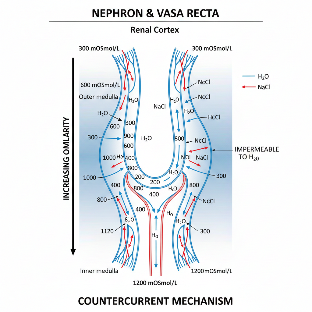
How does it pull this off? Through the genius of the Counter Current Mechanism. Notice this: the filtrate flows oppositely in the $\text{Henle's loop}$ and the blood flows oppositely in the $\text{vasa recta}$ (a 'U' shaped minute vessel running parallel to the loop). This opposite flow, combined with $\text{NaCl}$ and urea transport, maintains an increasing osmolarity gradient in the inner medullary interstitium. This gradient zooms from $300~mOsmolL^{-1}$ in the cortex to about $1200~mOsmolL^{-1}$ in the inner medulla. This steep gradient helps the collecting tubule pull out water easily, concentrating the final urine up to four times the initial filtrate.
NEET Checkpoint:
The key mechanism responsible for maintaining the $300$ to $1200~mOsmolL^{-1}$ gradient in the kidney medulla is the:
(a) Glomerular filtration
(b) Tubular secretion
(c) Counter Current Mechanism
(d) Micturition reflex
✅ Answer: (c) Counter Current Mechanism — The opposite flow in $\text{Henle's loop}$ and $\text{vasa recta}$ maintains this essential concentration gradient.
Figure below:
A clear diagram showing the nephron and vasa recta depicting the counter current mechanism and the increasing osmolarity gradient from $300~mOsmolL^{-1}$ to $1200~mOsmolL^{-1}$ towards the inner medulla, highlighting $\text{NaCl}$ and $\text{H}_2\text{O}$ movement.
Regulation of kidney function is a complex hormonal feedback loop involving the hypothalamus, JGA (Juxta Glomerular Apparatus), and the heart. The hypothalamus releases ADH/Vasopressin from the neurohypophysis. ADH facilitates water reabsorption from the latter parts of the tubule, preventing diuresis (water loss). If $\text{ADH}$ synthesis or release is impaired, you get Diabetes Insipidus, causing constant water loss and dehydration. Don't stress, yaar—it's easier than it looks!.
Now for the high-pressure mechanism: when glomerular blood flow or GFR falls, the $\text{JGA}$ cells release Renin. This triggers the Renin-Angiotensin Mechanism, leading to the production of Angiotensin II. Angiotensin II is a powerful vasoconstrictor that increases glomerular blood pressure and GFR. It also activates the adrenal cortex to release Aldosterone, which increases $\text{Na}^{+}$ and water reabsorption from the distal tubules, further boosting blood pressure and GFR.
NEET Checkpoint:
Angiotensin II acts as a powerful:
(a) Vasodilator
(b) Vasoconstrictor
(c) Diuretic
(d) Enzyme
✅ Answer: (b) Vasoconstrictor — Angiotensin II raises blood pressure by constricting blood vessels.
But wait, too much pressure is bad! When blood flow increases to the heart's atria, the heart releases Atrial Natriuretic Factor (ANF). ANF causes vasodilation and thereby decreases blood pressure, acting as a crucial check on the powerful renin-angiotensin mechanism.
Relatable Indian Example: Think of the $\text{Renin-Angiotensin-Aldosterone}$ system as a security guard who detects a pressure drop in the water pipes of an apartment building (the body). He quickly deploys a booster pump ($\text{Angiotensin II}$/$\text{Aldosterone}$) to increase the pressure. $\text{ANF}$ is the building supervisor who turns the booster pump down when the pressure goes too high, saving the pipes!
Kidneys don't work alone. Lungs remove vast amounts of $\text{CO}_2$ (approx. $200~mL/$ minute) and water. The Liver is key, excreting bile-containing substances like bilirubin, biliverdin, cholesterol, degraded steroid hormones, and drugs. The Skin excretes $\text{NaCl}$, urea, and lactic acid via sweat, and sterols and waxes via sebum.
Finally, the dark side: Disorders include Uremia (urea accumulation in blood due to kidney malfunction), which needs treatment via Dialysis (artificial kidney). Renal calculi are stones of crystallised salts (like oxalates) formed inside the kidney. Nephritis (specifically $\text{Glomerulonephritis}$) refers to the inflammation of the glomeruli of the kidney.
NEET Checkpoint:
Hemodialysis is a treatment method used specifically for which condition?
(a) Renal calculi
(b) Glomerulonephritis
(c) Uremia
(d) Diabetes Insipidus
✅ Answer: (c) Uremia — Hemodialysis removes accumulated urea from the blood when kidneys fail.
Practice Questions:
- A decrease in $GFR$ or blood pressure triggers the release of:
(a) $\text{ADH}$
(b) $\text{ANF}$
(c) $\text{Renin}$
(d) $\text{Aldosterone}$
✅ Explanation: A fall in $GFR$ activates $JG$ cells to release $\text{Renin}$, starting the cascade.
2. Which organ assists in excretion by eliminating $\text{bilirubin}$, $\text{biliverdin}$, and $\text{cholesterol}$?
(a) Lungs
(b) Skin
(c) Heart
(d) Liver
✅ Explanation: The $\text{Liver}$ secretes bile containing these substances for elimination.
Key Takeaway
Osmoregulation relies on the Counter Current Mechanism (Henle's loop/vasa recta) to maintain the $300$ to $1200~mOsmolL^{-1}$ gradient. $\text{ADH}$ promotes water conservation. The Renin-Angiotensin system increases $BP/GFR$, which is checked by the heart's ANF (vasodilation). Disorders like Uremia and Renal calculi affect kidney function.
10. Types of Movement and Muscle Structure: The Locomotion Funda
First, let’s talk movement types. Our human cells are busy bees exhibiting three main moves: amoeboid, ciliary, and muscular. The immune system's frontline soldiers, macrophages and leucocytes, use the amoeboid movement, shifting via pseudopodia formed by protoplasm streaming.
NEET Checkpoint:
Which cytoskeletal elements are involved in amoeboid movement?
(a) Microtubules
(b) Microfilaments
(c) Intermediate filaments
(d) None of these
✅ Answer: (b) Microfilaments — Cytoskeletal elements like microfilaments are involved in the movement of cells like macrophages.
The ciliary movement is key for internal plumbing. Imagine the coordinated sweep of tiny brushes: in your trachea, they help remove dust particles inhaled with air. Similarly, the passage of ova in the female reproductive tract is facilitated by ciliary action. Finally, there is the powerful muscular movement used for limbs, jaws, and the tongue, relying heavily on the muscle's contractile property.
Muscle Types: The Three Categories
Muscles are classified based on three criteria: location, appearance, and regulation. This classification is super important for NEET!
- Skeletal Muscles: Associated with bones, they look striated (striped) and are voluntary (you choose to move your arm).
- Visceral Muscles: Found in hollow organs (like the alimentary canal), they are smooth (non-striated) and involuntary (they work silently to transport food).
- Cardiac Muscles: Only found in the heart, these are striated but, critically, are involuntary.
Now, notice this funda: Skeletal muscle tissue is amazing because it possesses special properties: excitability, contractility, extensibility, and elasticity.
Imagine an auto-rickshaw stopping suddenly (a sudden external stimulus). Your ability to hold your balance involves the excitability and contractility of your skeletal muscles. Meanwhile, the muscles transporting your half-digested aloo tikki are the involuntary visceral type.
NEET Checkpoint:
The activities of visceral muscles are regulated by:
(a) Voluntary nervous control
(b) Involuntary neural control
(c) Only hormones
(d) Only stretch receptors
✅ Answer: (b) Involuntary neural control — Visceral muscle activities are not under the voluntary control of the nervous system.
Sarcomere: The Contraction Unit
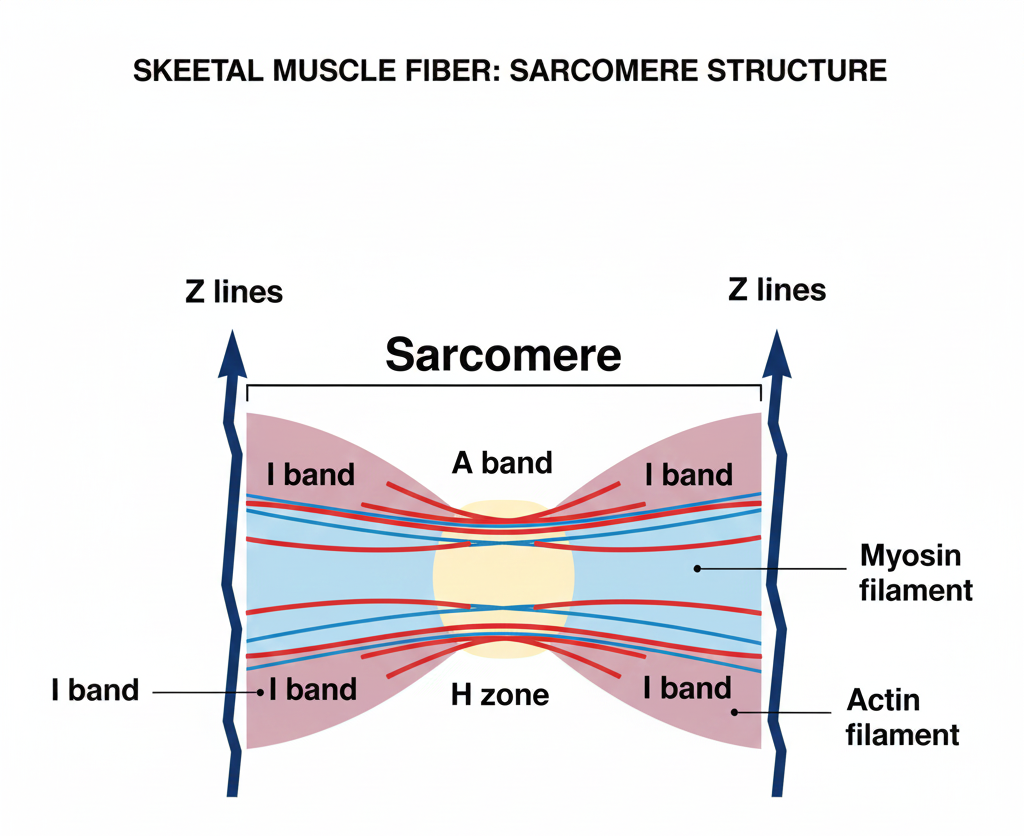
A muscle fibre contains numerous parallel filaments called myofibrils. These show alternating bands due to the arrangement of two proteins: Actin and Myosin.
The light band is the I-band (Isotropic band), which contains the thinner Actin filaments. The dark band is the A-band (Anisotropic band), which contains the thicker Myosin filaments.
The smallest functional unit of muscle contraction is the sarcomere. It’s defined as the segment of the myofibril between two successive '$Z$' lines. The '$Z$' line is an elastic fibre that bisects the '$I$' band. Here’s the trick: The central part of the '$A$' band, not overlapped by the thin Actin filaments in a resting state, is called the '$H$' zone.
NEET Checkpoint:
Which component defines the boundary of a single sarcomere?
(a) $A$ band
(b) $M$ line
(c) $Z$ line
(d) $H$ zone
✅ Answer: (c) $Z$ line — A sarcomere is the region between two successive '$Z$' lines.
Figure below:
A clear diagram showing the anatomy of a muscle fibre, highlighting two successive 'Z' lines, defining the sarcomere, and labelling the A band, I band, and H zone.
Practice Question:
The thick filament (Myosin) is found entirely within which band?
(a) I-band
(b) Z-line
(c) A-band
(d) H-zone
✅ Answer: (c) A-band — The dark A-band is where the thick Myosin filaments are located.
Key Takeaway
Movement Types: Amoeboid, Ciliary, Muscular.
Muscle Types: Skeletal (Striated, Voluntary), Visceral (Smooth, Involuntary), Cardiac (Striated, Involuntary).
Sarcomere: Functional unit between two successive '$Z$' lines. $I$-band contains Actin; $A$-band contains Myosin.
11. Mechanism of Muscle Contraction
Muscle contraction seems complex, but once you get the sliding filament theory clear, it's smoother than butter dosa! This theory explains that contraction happens when the thin filaments slide over the thick filaments. Notice this: the whole funda hinges on two major proteins: $\text{Actin}$ (thin) and $\text{Myosin}$ (thick).
Let's clear up the structure funda first. The thin $\text{Actin}$ filament is basically a polymer of monomeric 'G' (Globular) $\text{actins}$ forming two filamentous 'F' $\text{actins}$ wound helically. Associated with this are $\text{tropomyosin}$ and the complex protein $\text{Troponin}$. Here's the trick: in a resting muscle, a subunit of $\text{Troponin}$ cleverly masks the active binding sites for $\text{Myosin}$ on $\text{Actin}$.
Now for the bulky partner: the thick $\text{Myosin}$ filament is a polymer of $\text{Meromyosins}$. Each $\text{Meromyosin}$ has a heavy component called $\text{Heavy Meromyosin}$ ($\text{HMM}$), which is the globular head with a short arm, and a tail, the $\text{Light Meromyosin}$ ($\text{LMM}$). That globular head ($\text{HMM}$) is super important: it acts as an active $\text{ATPase}$ enzyme and has binding sites for both $\text{ATP}$ and $\text{Actin}$.
The Contraction Cycle
Contraction is initiated when a neural signal sent by the Central Nervous System ($\text{CNS}$) reaches the neuromuscular junction (or motor-end plate) via a motor neuron. This spot releases the neurotransmitter $\text{Acetyl choline}$, generating an action potential in the muscle's membrane ($\text{sarcolemma}$).
And that’s cool, right? This action potential causes a rapid release of $\text{calcium ions}$ ($\text{Ca}^{++}$) from the $\text{sarcoplasmic reticulum}$ into the $\text{sarcoplasm}$. This is the key activation step! The $\text{Ca}^{++}$ then binds to $\text{troponin}$, removing the masking from $\text{Actin}$’s active sites.
NEET Checkpoint:
The neurotransmitter released at the neuromuscular junction to initiate muscle contraction is:
(a) $\text{GABA}$
(b) $\text{Dopamine}$
(c) $\text{Acetyl choline}$
(d) $\text{Serotonin}$
✅ Answer: (c) $\text{Acetyl choline}$ — This specific neurotransmitter generates the action potential in the muscle fiber.
Utilising energy from $\text{ATP}$ hydrolysis, the $\text{Myosin}$ head binds to $\text{Actin}$, forming a cross bridge. This cross bridge acts like a strong arm pulling the $\text{Actin}$ filaments inward towards the centre of the '$\text{A}$ band'. This pulling action shortens the functional unit, the $\text{sarcomere}$. When the muscle shortens, the '$\text{I}$ bands' get reduced, while the '$\text{A bands}$' retain their length. Imagine pulling a heavy rickshaw towards you by repeatedly forming a grip, pulling, releasing the grip (breaking the cross bridge with new $\text{ATP}$), and repeating the pull (cross bridge formation and sliding).
NEET Checkpoint:
During muscle contraction, which structure remains unchanged in length?
(a) $\text{Sarcomere}$
(b) '$\text{I}$ band'
(c) '$\text{H}$ zone'
(d) '$\text{A}$ band'
✅ Answer: (d) '$\text{A}$ band' — The '$\text{A}$ band' retains its length, while the '$\text{I}$ bands' and the '$\text{H}$ zone' are reduced as the $\text{Actin}$ slides inward.
To stop the contraction (relaxation), the $\text{Ca}^{++}$ ions are pumped back into the $\text{sarcoplasmic cisternae}$, leading to the re-masking of the $\text{Actin}$ filaments.
Muscle Fibre Types: Red vs. White
Muscles aren't all the same, yaar. We classify them into two types based on their oxygen-storing pigment, $\text{myoglobin}$:
- Red fibres (Aerobic Muscles): These have a high $\text{myoglobin}$ content (hence reddish appearance) and plenty of mitochondria. They use the large amount of stored oxygen for aerobic $\text{ATP}$ production, meaning they are built for sustained activity.
- White fibres: These have very low $\text{myoglobin}$ (pale/whitish). They have fewer mitochondria but a high amount of $\text{sarcoplasmic reticulum}$. They rely on the anaerobic process for energy.
NEET Checkpoint:
The globular head of $\text{Myosin}$ is crucial because it contains active sites for $\text{Actin}$, $\text{ATP}$, and functions as an:
(a) $\text{Acetyl choline}$ receptor
(b) $\text{Ca}^{++}$ pump
(c) $\text{ATPase}$ enzyme
(d) $\text{Troponin}$ binder
✅ Answer: (c) $\text{ATPase}$ enzyme — The globular head ($\text{HMM}$) is specifically an active $\text{ATPase}$ enzyme, which hydrolyses $\text{ATP}$ to provide energy for cross-bridge movement.
Practice Questions :
- The critical event that removes the masking of active sites on $\text{Actin}$ filaments, allowing cross-bridge formation, is:
(a) Binding of $\text{Acetyl choline}$ to $\text{Troponin}$.
(b) Release of $\text{ADP}$ and $\text{P}_i$ from $\text{Myosin}$ head.
(c) $\text{Ca}^{++}$ binding to $\text{Troponin}$.
(d) Hydrolysis of $\text{ATP}$ by the $\text{Myosin}$ head.
✅ Explanation: The action potential triggers the release of $\text{Ca}^{++}$, which then binds to $\text{Troponin}$, moving the $\text{tropomyosin}$ to expose the active sites on $\text{Actin}$.
2. Compared to $\text{White fibres}$, $\text{Red fibres}$ typically possess:
(a) Low $\text{myoglobin}$ and few $\text{mitochondria}$.
(b) High $\text{sarcoplasmic reticulum}$ and anaerobic dependence.
(c) High $\text{myoglobin}$ and rich $\text{mitochondria}$.
(d) Lower $\text{ATPase}$ activity.
✅ Explanation: $\text{Red fibres}$ have high $\text{myoglobin}$ and plenty of $\text{mitochondria}$, making them aerobic and red-coloured.
Key Takeaway
The Mechanism of Muscle Contraction is governed by the Sliding Filament Theory. A neural impulse releases $\text{Ca}^{++}$ from the sarcoplasmic reticulum; $\text{Ca}^{++}$ binds to $\text{Troponin}$ to unmask $\text{Actin}$ sites. The $\text{Myosin}$ head (an $\text{ATPase}$) forms a cross-bridge and pulls $\text{Actin}$ inward, shortening the sarcomere. Muscles are classified into Red (aerobic, high $\text{myoglobin}$) and White (anaerobic, low $\text{myoglobin}$) fibres.
12. Skeletal System, Joints, and Musculoskeletal Disorders
Hey future doctor, let's talk about the funda of bones and movement—it’s the framework that literally holds you together! Our skeletal system consists of $206$ bones and a few cartilages. This amazing structure is primarily divided into two main categories: the Axial skeleton and the Appendicular skeleton.
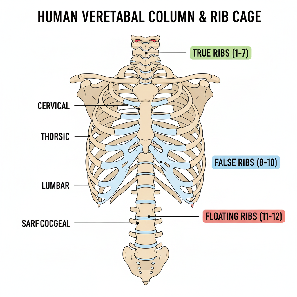
The Axial crew has $80$ bones and forms the central axis of the body, encompassing the skull, the vertebral column, the sternum, and the ribs. Now, notice this structure: the vertebral column has $26$ serially arranged units (vertebrae) and is differentiated into $7$ cervical, $12$ thoracic, $5$ lumbar, plus one fused sacral and one fused coccygeal region. The ribs, of which there are $12$ pairs, have a tricky classification: the first $7$ pairs are true ribs (they connect directly to the sternum), the $8^{th}$, $9^{th}$, and $10^{th}$ pairs are false ribs (they join the $7^{th}$ rib), and the last $2$ pairs are the floating ribs (they aren't connected ventrally at all). The Appendicular skeleton simply includes the bones of your limbs and their respective girdles.
Figure below:
A clear diagram showing the human vertebral column and rib cage, differentiating between the 7 true ribs, 3 false ribs, and 2 floating ribs.
NEET Checkpoint:
Which vertebral section consists of $12$ serially arranged units?
(a) Cervical
(b) Lumbar
(c) Thoracic
(d) Sacral
✅ Answer: (c) Thoracic — The thoracic region consists of $12$ vertebrae.
Joints are crucial—they are the points of contact between bones or between bones and cartilages. Think of joints as the hinges that allow voluntary movement! Structurally, we classify them into three major forms: Fibrous joints (like sutures in the skull) do not allow any movement. Cartilaginous joints (like those between adjacent vertebrae) permit limited movements. The most fun are the Synovial joints; they have a fluid-filled synovial cavity, allowing considerable movement (examples include the hinge joint, ball and socket joint, and pivot joint).
Relatable Indian Example: When you quickly turn your head to spot your tuition buddy calling you, the pivot joint between your atlas and axis vertebrae is doing the heavy lifting, allowing that rotational movement.
NEET Checkpoint:
The immovable joint type found in the skull is classified as:
(a) Synovial
(b) Pivot
(c) Cartilaginous
(d) Fibrous
✅ Answer: (d) Fibrous — These fuse end-to-end and do not allow any movement.
Now, sometimes the system has pangas. Muscular and skeletal disorders are vital to remember for NEET. Myasthenia gravis is an autoimmune disorder that attacks the neuromuscular junction, causing fatigue, weakness, and eventual paralysis of skeletal muscle. Tetany is characterized by rapid muscle spasms, often due to low $Ca^{++}$ concentration in the body fluid. A progressive degeneration of skeletal muscle, usually genetic, is known as Muscular dystrophy. Joint pain often points to Arthritis (inflammation of joints) or Gout (inflammation due to accumulation of uric acid crystals).
NEET Checkpoint:
Inflammation of joints resulting from the accumulation of uric acid crystals is known as: (a) Arthritis
(b) Tetany
(c) Muscular dystrophy
(d) Gout
✅ Answer: (d) Gout — This specific disorder is caused by uric acid crystal accumulation leading to joint inflammation.
Practice Questions:
1. Which set of ribs does not connect ventrally to the sternum or to another rib via cartilage?
(a) $7^{th}$ pair
(b) $8^{th}$, $9^{th}$, $10^{th}$ pairs
(c) $11^{th}$ and $12^{th}$ pairs
(d) $1^{st}$ pair
✅ Explanation: (c) The $11^{th}$ and $12^{th}$ pairs are the floating ribs, not connected ventrally.
2. The disorder involving fatigue, weakening, and paralysis of skeletal muscle, caused by an autoimmune response at the neuromuscular junction, is:
(a) Tetany
(b) Muscular dystrophy
(c) Myasthenia gravis
(d) Gout
✅ Explanation: Myasthenia gravis is the autoimmune disorder affecting the neuromuscular junction.
Key Takeaway
The human skeleton has $206$ bones, divided into Axial ($80$ bones) and Appendicular sections. Joints are structurally classified as Fibrous (immovable), Cartilaginous (limited movement, e.g., vertebrae), and Synovial (considerable movement, e.g., hinge/pivot). Disorders like Tetany (low $Ca^{++}$), Myasthenia gravis (autoimmune), and Gout (uric acid crystals) affect this system.
13. Neural System Organization and Impulse Generation
Hey NEET aspirant! Let's clear up the core funda of your nervous system—it's like the body's super-fast, fibre-optic network, coordinating everything.
The human neural system is neatly divided into two major parts, yaar: the Central Neural System ($\text{CNS}$) and the Peripheral Neural System ($\text{PNS}$). The $\text{CNS}$ is the main control room—it includes the brain and the spinal cord, handling all the major information processing. The $\text{PNS}$ consists of all the nerves that associate with the $\text{CNS}$ (the wiring that goes everywhere else).
NEET Checkpoint:
The site for information processing and control in the human body is the:
(a) PNS
(b) Autonomic System
(c) CNS
(d) Viscera
✅ Answer: (c) $\text{CNS}$ — Simple but important: the brain and spinal cord are the undisputed bosses!
The $\text{PNS}$ nerves have specific jobs. Think of them as a two-way courier service: Afferent fibres are the ones that take the impulses from the tissues/organs to the $\text{CNS}$. If you accidentally touch a hot tawa (griddle), the afferent nerves signal the danger to your brain. Efferent fibres carry regulatory impulses from the $\text{CNS}$ back to the peripheral tissues/organs to initiate a response (like pulling your hand away!).
The $\text{PNS}$ further splits into the Somatic Neural System (relays to skeletal muscles, controlling voluntary actions) and the Autonomic Neural System (relays to involuntary organs and smooth muscles). The Autonomic system is the true multi-tasker, splitting into the sympathetic and parasympathetic systems. Don't forget the Visceral nervous system; it’s a part of the $\text{PNS}$ that handles the complex network connecting the $\text{CNS}$ and the viscera (internal organs).
The Nerve Impulse Funda
The structural and functional unit of this entire system is the neuron. It has three main parts: the cell body, dendrites (which transmit impulses towards the cell body), and the axon (which transmits impulses away from the cell body).
Now, for the electrical magic! Neuronal membranes are "excitable" because they maintain a polarized state at rest—this is the resting potential. At rest, the membrane is more permeable to $\text{K}^{+}$ ions and nearly impermeable to $\text{Na}^{+}$ ions. Consequently, the axoplasm (inside) has a high concentration of $\text{K}^{+}$ and negatively charged proteins. The outside fluid has a high concentration of $\text{Na}^{+}$. This gradient is actively maintained by the sodium-potassium pump, which transports 3 $\text{Na}^{+}$ outwards for 2 $\text{K}^{+}$ into the cell. This makes the outer surface positive and the inner surface negative.
Figure below:
A clear diagram showing the depolarization wave along an axon, specifically illustrating the reversal of polarity (outer surface becoming negative, inner surface positive) at the stimulated site A, and the resultant current flow to the adjacent site B.
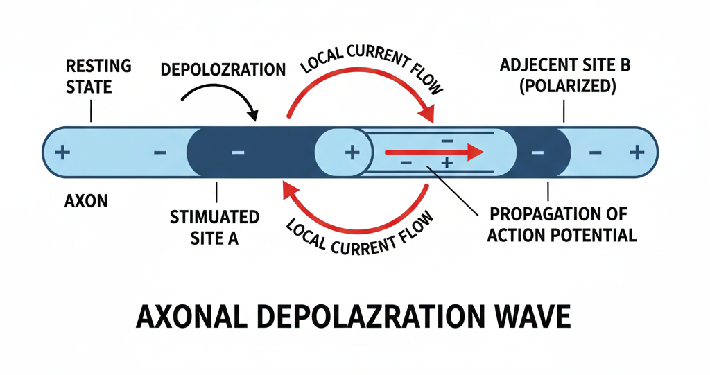
When a stimulus hits, shakti (power) is released! The membrane suddenly becomes freely permeable to $\text{Na}^{+}$. This causes a rapid influx of $\text{Na}^{+}$. This massive $\text{Na}^{+}$ entry reverses the polarity—the outside becomes negative and the inside positive—a process called depolarization. This instantaneous change creates the action potential (or nerve impulse), which is then conducted like a domino effect along the length of the axon.
NEET Checkpoint:
During the resting state of a neural membrane, the axoplasm contains high concentrations of: (a) $\text{Na}^{+}$ and $\text{K}^{+}$
(b) $\text{Na}^{+}$ and positive proteins
(c) $\text{K}^{+}$ and negatively charged proteins
(d) $\text{Na}^{+}$ only
✅ Answer: (c) $\text{K}^{+}$ and negatively charged proteins — The pump keeps $\text{Na}^{+}$ low inside while retaining $\text{K}^{+}$ and fixed negative proteins.
Practice Questions:
- Which structure transmits nerve impulses away from the neuron’s cell body?
(a) Nissl’s granules
(b) Dendrite
(c) Axon
(d) Synaptic knob
✅ Answer: (c) Axon — Dendrites carry impulses towards the cell body, while the axon carries them away.
2. The action potential (nerve impulse) is fundamentally characterized by:
(a) $\text{K}^{+}$ efflux
(b) $\text{Na}^{+}$ influx
(c) $\text{Na}^{+}$ efflux
(d) $\text{K}^{+}$ influx
✅ Answer: (b) $\text{Na}^{+}$ influx — The rapid entry (influx) of $\text{Na}^{+}$ ions reverses the polarity, causing depolarization.
NEET Checkpoint:
The ratio of ions transported by the $\text{Na}^{+}-\text{K}^{+}$ pump is:
(a) $2 \text{Na}^{+}$ out for $3 \text{K}^{+}$ in
(b) $3 \text{Na}^{+}$ out for $2 \text{K}^{+}$ in
(c) $1 \text{Na}^{+}$ out for $1 \text{K}^{+}$ in
(d) $3 \text{Na}^{+}$ out for $3 \text{K}^{+}$ in
✅ Answer: (b) $3 \text{Na}^{+}$ out for $2 \text{K}^{+}$ in — Remember the magic ratio: three sodium ions are pushed out for every two potassium ions brought in, maintaining that crucial resting potential.
Key Takeaway
The nervous system hierarchy moves from $\text{CNS}$ (control) to $\text{PNS}$ (nerves). Neurons maintain a polarized resting potential (positive outside, negative inside) using the $3 \text{Na}^{+}$ out / $2 \text{K}^{+}$ in pump. An impulse is generated by rapid $\text{Na}^{+}$ influx, leading to temporary depolarization (the action potential).
14. Impulse Transmission and Central Neural System Structure
Don't stress, yaar—the nervous system is just a sophisticated communication network! We’re going to cover how signals jump between neurons and then meet the Big Boss: your brain.
The Synapse: Where Neurons Chat
A nerve impulse is transmitted from one neuron to the next through junctions called synapses. A synapse is essentially formed by the membranes of a pre-synaptic neuron and a post-synaptic neuron.
These junctions come in two flavours:
1. Electrical Synapses: Here, the pre- and post-synaptic membranes are in very close proximity. This allows electrical current to flow directly from one neuron to the other—like a high-speed dedicated line! Transmission across electrical synapses is always faster than chemical ones, but hey, they are rare in our system.
NEET Checkpoint:
Transmission across which type of synapse is very similar to impulse conduction along a single axon?
(A) Chemical synapse
(B) Mixed synapse
(C) Gap junction synapse
(D) Electrical synapse
✅ Answer: (D) Electrical synapse — Because current flows directly between the membranes.
2. Chemical Synapses: These are the common ones, where the two membranes are separated by a fluid-filled space called the synaptic cleft. When an action potential arrives at the axon terminal, it stimulates vesicles filled with neurotransmitters (NTs) to fuse with the membrane and release those chemicals into the synaptic cleft. These NTs then bind to specific receptors on the post-synaptic membrane, generating a new potential (which can be either excitatory or inhibitory). Think of neurotransmitters as the vital ingredients needed to pass the signal.
CNS: The Command and Control Centre
The brain is the 'command and control system' and the central information processing organ of our body. It handles everything from voluntary movement and body balance to thermoregulation, memory, and even your emotions and thoughts.
This precious organ is well-protected! It sits inside the skull and is covered by three layers of cranial meninges:
1. Outer layer: dura mater
2. Middle thin layer: arachnoid
3. Inner layer: pia mater (in contact with brain tissue)
Figure below:
A clear sagittal section diagram of the human brain highlighting the forebrain (cerebrum, thalamus, hypothalamus), midbrain, and hindbrain (pons, cerebellum, medulla).
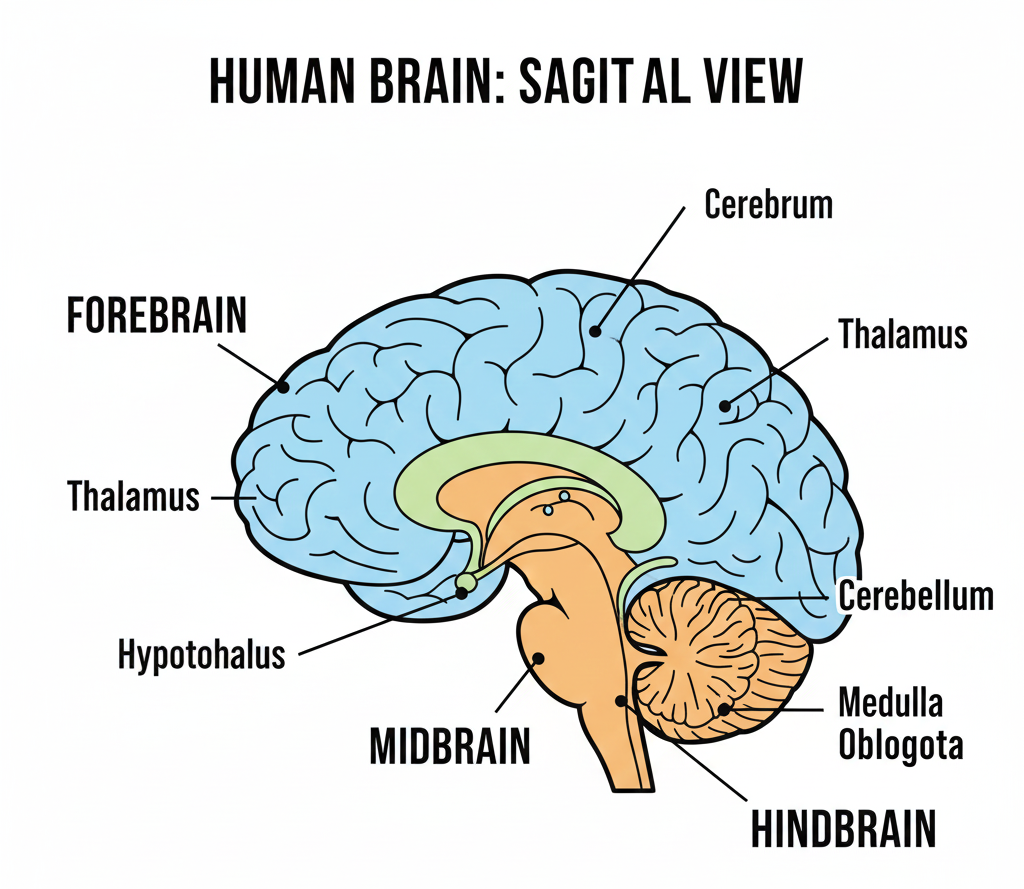
The brain itself is divided into three major parts: forebrain, midbrain, and hindbrain.
NEET Checkpoint:
Which meningeal layer is in direct contact with the brain tissue?
(A) Dura mater
(B) Arachnoid mater
(C) Pia mater
(D) Epidermis
✅ Answer: (C) Pia mater — It is the innermost, delicate layer.
Forebrain: The Think Tank
The Forebrain is composed of the cerebrum, thalamus, and hypothalamus.
- Cerebrum: This is the largest part, longitudinally split into two hemispheres connected by a tract of nerve fibres called the corpus callosum. It is the site for complex features handled by association areas, like intersensory associations, memory, and communication. Imagine the corpus callosum as the biggest national highway, ensuring the two halves of your brain talk seamlessly!
- Thalamus: The cerebrum wraps around this structure, which acts as a major coordinating centre for sensory and motor signalling.
- Hypothalamus: Located at the base of the thalamus, it contains several centres that control body temperature and the urge for eating and drinking. It also secretes hypothalamic hormones.
Hindbrain: The Vitals Keeper
The Hindbrain comprises the pons, cerebellum, and medulla.
- Pons: Consists of fibre tracts that interconnect different regions of the brain.
- Cerebellum: This looks intricate because it has a very convoluted surface to provide additional space for a massive number of neurons. It’s key for integrating sensory information and movement.
- Medulla (Medulla Oblongata): Connected to the spinal cord, the medulla controls vital involuntary functions such as respiration, cardiovascular reflexes, and gastric secretions.
NEET Checkpoint:
Which brain region contains centres that regulate respiration and cardiovascular reflexes?
(A) Cerebrum
(B) Cerebellum
(C) Pons
(D) Medulla oblongata
✅ Answer: (D) Medulla — It controls these vital involuntary organ functions.
Practice Questions:
- The tract of nerve fibres connecting the two cerebral hemispheres is called the:
(a) Thalamus
(b) Corpus callosum
(c) Pons
(d) Arachnoid
✅ Answer: (b) Corpus callosum — This structure connects the left and right cerebral hemispheres.
2. At a chemical synapse, the membranes of pre- and post-synaptic neurons are separated by the:
(a) Myelin sheath
(b) Node of Ranvier
(c) Synaptic knob
(d) Synaptic cleft
✅ Answer: (d) Synaptic cleft — This fluid-filled space is characteristic of chemical synapses, across which neurotransmitters travel.
Key Takeaway
Impulse Transmission primarily occurs at synapses, which are either fast electrical or neurotransmitter-dependent chemical junctions. The brain is protected by the skull and three meninges. Key parts include the Forebrain (Cerebrum for higher function/memory; Thalamus for coordination; Hypothalamus for temperature/thirst) and the Hindbrain (Pons interconnects; Cerebellum has a convoluted surface; Medulla controls vital reflexes like respiration).
15. Chemical Coordination, Endocrine Glands, and Disorders
Hey junior! Let’s talk about the body’s "wireless" communication system—the endocrine system. Unlike the nervous system's high-speed wires, this system uses hormones: non-nutrient, intercellular messengers produced in trace amounts by ductless glands.
Think of the Hypothalamus as the CEO of a company; it sits at the base of the forebrain and sends "releasing" or "inhibiting" memos to the Pituitary (the manager) to control everything from growth to stress. The Pituitary then releases hormones like GH (Growth Hormone), TSH, and ADH. For instance, if your GH levels go crazy in adulthood, you might end up with Acromegaly—a condition where facial features become severely disfigured.
Hypothalamus–Pituitary Axis
The hypothalamus serves as the principal link between the nervous and endocrine systems. Located at the base of the forebrain, it integrates neural inputs and translates them into endocrine signals. It regulates the pituitary gland through releasing hormones (which stimulate pituitary secretion) and inhibiting hormones (which suppress secretion). For example, Gonadotropin Releasing Hormone (GnRH) controls the release of LH and FSH, while dopamine acts as a prolactin-inhibiting hormone.
The pituitary gland, often referred to as the master gland, lies just below the hypothalamus. The anterior pituitary synthesizes and secretes trophic hormones such as Growth Hormone (GH), Thyroid Stimulating Hormone (TSH), Adrenocorticotropic Hormone (ACTH), Follicle Stimulating Hormone (FSH), Luteinizing Hormone (LH), and Prolactin. These hormones regulate other endocrine glands or physiological processes.
The posterior pituitary does not synthesize hormones but stores and releases Oxytocin and Antidiuretic Hormone (ADH or Vasopressin), both of which are synthesized in the hypothalamus. ADH plays a crucial role in maintaining water balance by increasing water reabsorption in the kidneys, thereby preventing excessive urine formation.
Pineal Gland and Biological Rhythm
The pineal gland, situated deep within the brain, secretes melatonin, a hormone responsible for regulating the body’s diurnal or circadian rhythm. Melatonin influences sleep–wake cycles, seasonal reproductive rhythms, and timing of puberty. Its secretion is higher during darkness and decreases in light, acting as the body’s internal clock.
NEET Checkpoint:
Which hormone is responsible for maintaining the 24-hour (diurnal) rhythm of our body?
(a) Thyroxine
(b) Melatonin
(c) Insulin
(d) Adrenaline
✅ Answer: (b) Melatonin — Secreted by the Pineal gland, it acts as your internal alarm clock for sleep-wake cycles.
In the "emergency ward," we have Adrenaline and Noradrenaline from the adrenal medulla. They are your "fight or flight" buddies—kind of like the rush you feel when a stray dog starts chasing your cycle in the colony! They increase your heart beat and alertness instantly.
NEET Checkpoint:
Which hormone acts as a "hypercalcemic" factor?
(a) Thyrocalcitonin (TCT)
(b) Parathyroid Hormone (PTH)
(c) Cortisol
(d) Glucagon
✅ Answer: (b) PTH — It increases blood Ca2+ levels by stimulating bone resorption.
Hormones don't just work randomly. Peptide hormones (like Insulin) bind to membrane-bound receptors and generate second messengers like $cAMP$ or $Ca^{++}$. Steroid hormones, however, usually enter the cell to interact with intracellular receptors and regulate your genes directly.
Even organs not classified as major endocrine glands act as secret agents! The Heart's atrial wall releases ANF (Atrial Natriuretic Factor) when blood pressure (BP) increases, causing vasodilation to reduce BP. The Kidney produces Erythropoietin which stimulates erythropoiesis (RBC formation).
NEET Checkpoint:
Underproduction of hormones by the adrenal cortex leads to:
(a) Grave’s disease
(b) Diabetes mellitus
(c) Addison’s disease
(d) Acromegaly
✅ Answer: (c) Addison’s disease — This causes acute weakness and fatigue due to altered carbohydrate metabolism.
Here's the fundamental funda: Endocrine glands are ductless glands. Their secretions, hormones, are defined as non-nutrient chemicals acting as intercellular messengers produced in trace amounts.
The main boss is the Hypothalamus, regulating the Pituitary via two types of chemical signals: releasing hormones (which stimulate secretion) and inhibiting hormones (which suppress it). The Pituitary gland, often called the master gland, pumps out six trophic hormones from its anterior part, including GH (Growth Hormone), TSH, LH, and FSH. Notice this: the Posterior Pituitary just stores Oxytocin and Vasopressin (or ADH) that the hypothalamus actually synthesizes. $ADH$ helps conserve water by stimulating reabsorption in the kidney, thereby preventing diuresis.
Now for some key players. The Pancreas secretes Insulin (the hypoglycemic hormone, which lowers blood glucose by enhancing cellular uptake) and Glucagon (the hyperglycemic hormone, which raises blood glucose). The Adrenal medulla blasts out Adrenaline (Catecholamines), famously known as the emergency hormones or hormones of "Fight or Flight".
How do hormones work their magic? They bind to specific receptors. Peptide hormones (like Insulin) bind to membrane-bound receptors and generally use second messengers (like cyclic $AMP$ or $Ca^{++}$) to transmit the signal inside. In contrast, Steroid hormones utilize intracellular receptors to form a hormone-receptor complex that directly interacts with the genome, regulating gene expression. Imagine trying to unlock your phone with the wrong fingerprint—it won't open unless the specific receptor matches!
Disorders Alert! Deficiency of Insulin leads to Diabetes Mellitus (marked by hyperglycemia and ketone bodies). Excess $GH$ secretion in adults causes severe disfigurement called Acromegaly. Cretinism (stunted growth, mental retardation) results from hypothyroidism during pregnancy. Addison’s disease is caused by the underproduction of adrenal cortex hormones, leading to acute weakness.
Thyroid Gland and Metabolism
The thyroid gland produces Triiodothyronine (T₃) and Thyroxine (T₄), hormones essential for regulating basal metabolic rate (BMR), oxygen consumption, and overall metabolism of carbohydrates, proteins, and fats. Adequate iodine intake is necessary for their synthesis. Deficiency of thyroid hormones during infancy leads to cretinism, characterized by stunted growth and mental retardation, whereas deficiency in adults causes myxedema. Enlargement of the thyroid gland due to iodine deficiency results in goitre.
If things go wrong, you might face Goitre (enlargement) or Cretinism in babies. Meanwhile, the Pancreas acts as a dual agent, with Insulin lowering blood glucose and Glucagon raising it. Even your heart has a side hustle, secreting ANF to decrease blood pressure.
Practice Question:
1. Hyposecretion of $GH$ during childhood results in:
(a) Acromegaly
(b) Gigantism
(c) Pituitary Dwarfism
(d) Addison's disease
✅ Answer: (c) Pituitary Dwarfism — Low secretion of GH results in stunted growth, leading to pituitary dwarfism.
2. Cretinism in children is often a result of:
(a) Hyperthyroidism
(b) Hypothyroidism during pregnancy
(c) Excess GH
(d) Insulin deficiency.
✅ Answer: (b) Hypothyroidism during pregnancy — It leads to stunted growth and mental retardation.
Mechanism of Hormone Action
Hormones exert their effects by binding to specific receptors present on or within target cells. Peptide and protein hormones, being water-soluble, bind to membrane-bound receptors and act through second messengers such as cyclic AMP (cAMP) or calcium ions. These pathways amplify the hormonal signal inside the cell.
In contrast, steroid hormones are lipid-soluble and diffuse through the cell membrane to bind intracellular receptors. The hormone–receptor complex directly interacts with DNA, regulating gene transcription and protein synthesis. This results in slower but longer-lasting effects.
Feedback Regulation
Hormone secretion is regulated mainly by negative feedback mechanisms, where increased levels of a hormone inhibit its further release. For example, high levels of thyroid hormones suppress TSH secretion. This maintains hormonal balance and homeostasis within the body.
Key Takeaway
• Hormones: Non-nutrient, trace intercellular messengers.
• Emergency Hormones: Adrenaline/Noradrenaline (Fight or Flight).
• Mechanism: Peptide hormones use second messengers (e.g., cAMP); Steroids regulate gene expression
The action mechanism depends on the hormone's chemical nature: Peptide hormones use membrane receptors/second messengers ($cAMP$) while Steroid hormones use intracellular receptors to modify gene expression. Key regulators include ANF (lowers $BP$), Erythropoietin (stimulates $RBC$ formation), and Insulin/Glucagon (blood sugar control).