1. The Fundamental Unit of Life
Listen up, junior! Ever wondered what makes a living thing actually "alive"? It's not just about breathing or moving; it’s all about the cell. Think of it like a cricket stadium: the stadium is the organism, but without the individual bricks (the cells), you’ve got nothing but an empty field!.
A single cell is a total powerhouse. Unicellular organisms like bacteria can live completely on their own and perform every essential life function. In biology, "less is more" doesn't work—anything less than a complete cell cannot ensure independent living.
NEET Checkpoint:
Who was the first person to see and describe a live cell?
(a) Robert Brown
(b) Robert Hooke
(c) Antonie von Leeuwenhoek
(d) Rudolf Virchow
✅ Answer: (c) Antonie von Leeuwenhoek — Robert Brown came much later to find the nucleus in 1831!.
The Cell Theory was a team effort. In 1838, Matthias Schleiden (a botanist) noticed all plants are made of cells. Then, in 1839, Theodore Schwann (a zoologist) found that animal cells have a thin outer layer we now call the plasma membrane. Schwann also made a "googly" discovery: plant cells are unique because they have a cell wall.
NEET Checkpoint:
Which scientist proposed that the bodies of animals and plants are composed of cells and products of cells?
(a) Robert Hooke
(b) Virchow
(c) Schleiden and Schwann
(d) Leeuwenhoek
✅ Answer: (b) Schleiden and Schwann — He was the one who linked the two worlds together in 1839!.
But wait, there was a missing piece! They didn't know where new cells came from. Enter Rudolf Virchow (1855). He dropped the famous line: Omnis cellula-e cellula, meaning all cells arise from pre-existing cells.
NEET Checkpoint:
What was Rudolf Virchow’s major contribution to the Cell Theory?
(a) Discovery of the cell
(b) Cells are the basic unit of life
(c) All cells arise from pre-existing cells
(d) Cells contain genetic material
✅ Answer: (c) He explained that cells divide and new cells are formed from pre-existing cells, giving the theory its final shape.
Practice Questions:
1. According to the modern cell theory, which of the following is true?
(a) All cells are formed de novo.
(b) All living organisms are composed of cells and products of cells.
(c) Only multicellular organisms are made of cells.
(d) Cells are not the functional unit of life.
✅ Answer: (b) — This is the core "funda" of the modified theory by Schleiden, Schwann, and Virchow.
2. Which of these is a unique characteristic of plant cells according to Schwann's studies?
(a) Presence of Nucleus
(b) Presence of Plasma membrane
(c) Presence of Cell wall
(d) Presence of Cytoplasm
✅ Answer: (c) — Schwann concluded that the cell wall is a unique character of plant cells.
Key Takeaway
- Cell: The fundamental structural and functional unit of all living organisms.
- Leeuwenhoek: Saw live cells; Robert Brown: Discovered the nucleus.
- Modern Cell Theory: All organisms are made of cells, and all cells arise from pre-existing cells (Omnis cellula-e cellula).
2. Prokaryotic Cell Structure
Listen up, junior! If the biological world were a tech market, prokaryotes would be the rugged, "no-nonsense" base models. They don't have fancy compartments, but they are incredibly efficient. This squad includes bacteria, blue-green algae, mycoplasma, and PPLO (Pleuro Pneumonia Like Organisms).
NEET Checkpoint:
Which is the smallest cell mentioned in your NCERT?
(a) Bacteria
(b) PPLO
(c) Nerve cell
(d) RBC
✅ Answer: (b) PPLO — At about $0.1~\mu m$, they make even normal bacteria look huge!.
Now, let’s clear up the cell envelope funda. It’s a three-layered security system: the outermost glycocalyx, the middle cell wall (for shape and support), and the innermost plasma membrane. Think of it like a Triple-Layered Cheese Burst Pizza: the crust is the cell wall, the sticky cheese on top is the glycocalyx, and the thin base holding it all together is the plasma membrane.
NEET Checkpoint:
What do we call a thick and tough glycocalyx?
(a) Cell wall
(b) Slime layer
(c) Capsule
(d) Plasma membrane
✅ Answer: (c) A capsule. If it's just a loose sheath, it's a slime layer.
Inside, the genetic material is "naked"—no nuclear membrane here, yaar! You’ll find a single circular chromosome (genomic DNA) and often small extra-circular DNA called plasmids. These plasmids are like "bonus cheat codes" that give bacteria cool traits like antibiotic resistance.
For the "gadgets," prokaryotes have mesosomes—clever infoldings of the plasma membrane that help with respiration, secretion, and DNA replication. If the bacterium is motile, it uses a flagellum (made of a filament, hook, and basal body) to zoom around.
NEET Checkpoint:
Are ribosomes found in prokaryotes?
(a) No, ribosomes are absent
(b) Only attached ribosomes are present
(c) Yes, free ribosomes are present
(d) Only 80S ribosomes are present
✅ Answer: (c) Yes! They are $70S$ type, and several can attach to a single mRNA to form a polysome.
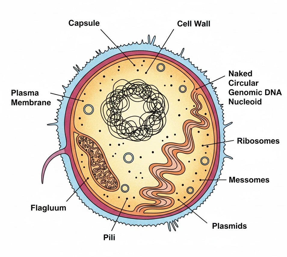
Figure below:
A detailed diagram of a prokaryotic cell showing the three-layered envelope, naked circular genomic DNA, small plasmids, and the infolded mesosomes.
Practice Questions:
1. Which structure is an extension of the plasma membrane into the cell?
(a) Flagella
(b) Mesosome
(c) Pili
(d) Fimbriae
✅ Answer: (b) — Mesosomes are specialized infoldings for vital functions like respiration.
2. Plasmids in bacteria confer which of the following?
(a) Mobility
(b) Protein synthesis
(c) Antibiotic resistance
(d) Cell wall formation
✅ Answer: (c) — These small circular DNA molecules provide unique phenotypic characters.
Key Takeaway
- Squad: Bacteria, BGA, Mycoplasma, PPLO.
- Envelope: Glycocalyx $\rightarrow$ Cell Wall $\rightarrow$ Plasma Membrane.
- DNA: Naked genomic DNA + Plasmids (for antibiotic resistance).
- Specialities: Mesosomes (respiration) and Flagella (motility).
3. Eukaryotic Cells & Comparison
Hey there, future doctor! Ready to level up? While prokaryotes are like basic studio apartments, eukaryotic cells are like luxury bungalows with multiple rooms. This "room-wise" arrangement is what we call extensive compartmentalisation, thanks to membrane-bound organelles.
The cytoplasm is the heart of the action—think of it as the main cricket pitch (the main arena) where all the metabolic "sixers" and chemical reactions happen to keep the cell in a 'living state'.
NEET Checkpoint:
Which part of the cell is considered the 'main arena' for cellular activities?
(a) Nucleus
(b) Mitochondria
(c) Endoplasmic reticulum
(d) Cytoplasm
✅ Answer: (d) Cytoplasm — It’s where all the chemical dhamaal happens to keep the cell alive!
Now, let's clear the funda on Plant vs. Animal cells. Plant cells are like traditional Indian homes with a sturdy char-diwari (cell wall) and a massive storage room (large central vacuole) for all the extra rations. They also have plastids to make food. Animal cells are more flexible; they lack those walls but have a special "security detail" called centrioles to help in cell division.
NEET Checkpoint:
Name an organelle found in animal cells but absent in almost all plant cells.
(a) Mitochondria
(b) Golgi apparatus
(c) Ribosomes
(d) Centrioles
✅ Answer: Centrioles — These help our animal cells divide properly during mitosis!
NEET Checkpoint:
Which structures are unique to plant cells compared to animal cells?
(a) Mitochondria and ribosomes
(b) Nucleus and endoplasmic reticulum
(c) Cell wall, plastids, and large central vacuole
(d) Lysosomes and centrioles
✅ Answer: (c) Cell walls, plastids, and a large central vacuole.
Notice this: even though they look different, both have their genetic material organised into chromosomes inside a proper nucleus.
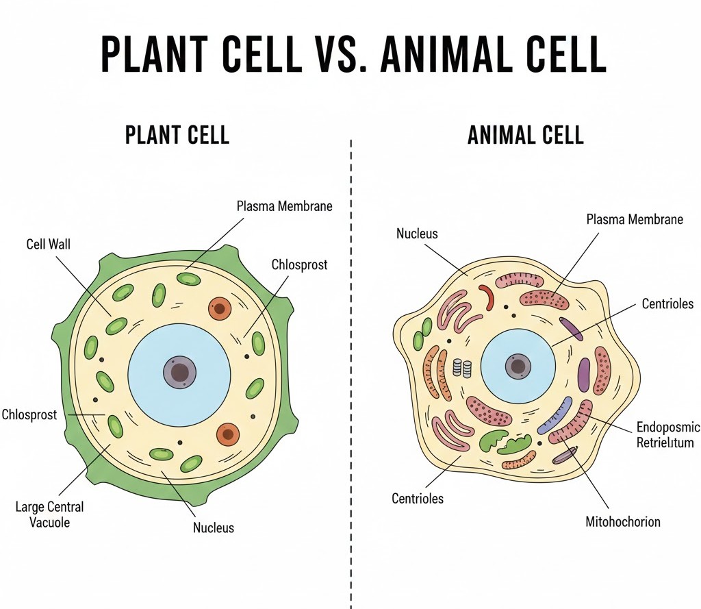
Figure below:
A clear, side-by-side comparative diagram of a typical Plant Cell showing a large central vacuole, cell wall, and chloroplasts, and an Animal Cell showing centrioles and a more flexible boundary.
Practice Question:
Which of the following is present in a plant cell but absent in an animal cell?
(a) Mitochondria
(b) Ribosomes
(c) Plastids
(d) Cytoplasm
✅ Answer: (c) — Plants need plastids (like chloroplasts) for photosynthesis, which animals don't do!
Don't stress, yaar—just remember that plants have the "big three" (Wall, Plastid, Vacuole) and you're golden! Now that you've got this, wait till you see how the Cell Membrane actually controls who enters the party.
Key Takeaway
- Eukaryotes: High compartmentalisation via organelles.
- Cytoplasm: The main arena of cellular activity.
- Plants: Have Cell Wall, Plastids, and Large Vacuoles.
- Animals: Have Centrioles; lack Cell Walls.
4. Cell Membrane & Cell Wall
Listen up, champ! Every cell needs a solid "security system" and a sturdy "boundary wall." That’s exactly what the Cell Membrane and Cell Wall do. In 1972, Singer and Nicolson gave us the Fluid Mosaic Model, which is the gold standard for understanding the membrane. Imagine a sea of phospholipids where proteins are like icebergs floating around; this quasi-fluid nature allows proteins to move laterally, which is vital for functions like cell growth and secretion.
The membrane is a bilayer of phospholipids with "polar heads" facing the outside and "hydrophobic tails" tucked safely inside to avoid water. It also contains cholesterol to keep things stable.
Example: Think of a crowded Mumbai local train. The "selectively permeable" membrane is like the door—only those with the right "ticket" (specific molecules) get to pass through easily!
NEET Checkpoint:
What is the "glue" that holds neighbouring plant cells together?
(a) Cell wall
(b) Calcium pectate
(c) Middle lamella
(d) Plasma membrane
✅ Answer: (c) Middle lamella — It is a layer mainly made of calcium pectate.
When it comes to Membrane Transport, it’s all about the energy, yaar! Passive transport (like neutral solutes moving by diffusion or water by osmosis) happens along the concentration gradient without spending a single calorie. But for the tough stuff, we use Active transport, like the $Na^{+}/K^{+}$ pump, which gulps down ATP to move molecules against the gradient.
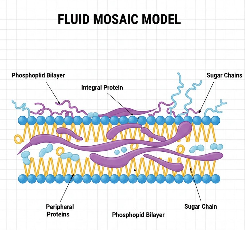
Figure below:
A detailed diagram of the Fluid Mosaic Model showing the phospholipid bilayer, integral and peripheral proteins, cholesterol, and sugar chains.
NEET Checkpoint:
Which molecules require "carrier proteins" to cross the nonpolar lipid bilayer?
(a) Non-polar molecules
(b) Gases like O₂ and CO₂
(c) Polar molecules
(d) Lipid-soluble molecules
✅ Answer: (c) Polar molecules — Because they cannot pass through the nonpolar tail region of the membrane.
Now, don't confuse the membrane with the Cell Wall. The wall is a rigid, non-living outer covering found in plants and fungi. In algae, it’s made of cellulose, galactans, and even calcium carbonate $CaCO_{3}$, while higher plants use cellulose, hemicellulose, and pectins.
Humour: The cell wall is like that strict colony watchman—it gives shape and protects the cell from "mechanical damage" and unwanted guests (infections)!
NEET Checkpoint:
Where is the secondary cell wall formed in a maturing plant cell?
(a) Outside the middle lamella
(b) Between middle lamella and primary wall
(c) On the inner side of the primary cell wall
(d) Outside the plasma membrane
✅ Answer: (c) On the inner side (towards the membrane) of the cell.
Practice Questions:
1. The fluid nature of the membrane is important for which of the following?
(a) Cell division
(b) Endocytosis
(c) Intercellular junctions
(d) All of these
✅ Answer: (d) — The sources state fluidity is essential for all these functions.
2. Which of the following is a characteristic of active transport?
(a) Occurs along the concentration gradient
(b) No energy required
(c) Uses ATP
(d) Simple diffusion
✅ Answer: (c) — Active transport is an energy-dependent process using ATP.
Key Takeaway
- Membrane: Phospholipid bilayer + Proteins + Cholesterol; semi-permeable.
- Transport: Passive (No ATP) vs. Active (Uses ATP, e.g., $Na^{+}/K^{+}$ pump).
- Cell Wall: Cellulose-based in plants; contains $CaCO_{3}$ in algae.
- Middle Lamella: Made of calcium pectate.
5. Endomembrane System: ER & Golgi
Welcome to the cell's very own logistics department! The Endomembrane System isn't just a random collection; it’s a coordinated team consisting of the Endoplasmic Reticulum (ER), Golgi complex, lysosomes, and vacuoles. Notice this: mitochondria and chloroplasts are solo players and are NOT considered part of this system because their functions aren't coordinated with these guys. Let's clear up this funda: every team needs a factory and a courier service, right?.
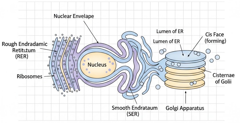
Think of the ER as the vast factory floor. It is a network of tiny tubular structures that divides the intracellular space into the luminal (inside ER) and extra-luminal (cytoplasm) compartments. We have two main departments here: Rough ER (RER), which is studded with ribosomes for protein synthesis and secretion, and Smooth ER (SER), the specialist for lipid synthesis and steroidal hormones in animal cells.
NEET Checkpoint:
Where are steroidal hormones synthesised in animal cells?
(a) RER
(b) SER
(c) Golgi
(d) Lysosomes
✅ Answer: (b) SER — It's the smooth operator for lipids and steroids!
Next is the Golgi Apparatus, first seen by Camillo Golgi in 1898. It consists of many flat, disc-shaped sacs called cisternae (about $0.5~\mu m$ to $1.0~\mu m$ in diameter) stacked parallel to each other. It has a convex cis (forming) face to receive materials and a concave trans (maturing) face to ship them out. Even the cell needs a reliable courier service for its glycoproteins and glycolipids!.
NEET Checkpoint:
The maturing face of the Golgi apparatus is:
(a) Cis
(b) Convex
(c) Trans
(d) Nucleus-facing
✅ Answer: (c) Trans — Also known as the concave face where stuff gets ready to leave!
Relatable Example: Imagine a swiggy delivery kitchen. The RER/SER is the chef cooking the food (proteins/lipids), while the Golgi is the packing guy who puts it in boxes, labels them (modifications like glycoproteins), and hands them to the driver at the trans exit!
NEET Checkpoint:
Which organelle is the important site for the formation of glycoproteins?
(a) Mitochondria
(b) Golgi apparatus
(c) Ribosomes
(d) Lysosomes
✅ Answer: (b) Golgi apparatus — It adds the "sugar" tag to your proteins!
Figure below:
A clear diagram showing the Endoplasmic Reticulum with ribosomes (RER) and without (SER) connected to the nuclear envelope, alongside the stacked cisternae of the Golgi apparatus showing the distinct cis and trans faces.
Practice Question:
Materials to be packaged in the form of vesicles from the ER fuse with which part of the Golgi?
(a) Trans face
(b) Cisternae
(c) Cis face
(d) Maturing face
✅ Answer: (c) — Materials always enter the "forming" or cis face first!
Key Takeaway
- Endomembrane System: Coordinated group of ER, Golgi, Lysosomes, and Vacuoles.
- RER: Protein synthesis; SER: Lipid synthesis.
- Golgi: Packaging center; modifies proteins into glycoproteins; uses cis (entry) and trans (exit) faces.
6. Endomembrane System: Lysosomes & Vacuoles
Listen up, junior! Now that we’ve seen the "post office" (Golgi), let’s look at the cell’s waste management and storage units. Don't stress, yaar—this funda is easier than it looks!
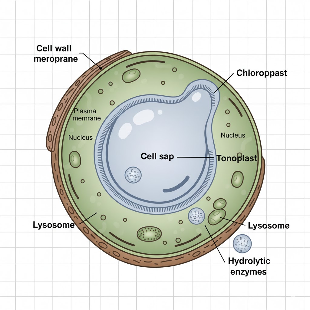
Lysosomes are membrane-bound vesicles formed right inside the Golgi apparatus. Think of them as the cell's "suicide squads" or recycling bins. They are packed with hydrolytic enzymes (hydrolases) like lipases, proteases, carbohydrases, and nucleases. These guys are only "active" when the environment is acidic—much like how a student only gets active right before the exams! These enzymes are total pros at digesting carbohydrates, proteins, lipids, and even nucleic acids.
NEET Checkpoint:
At what $pH$ are lysosomal enzymes most active?
(a) Neutral pH
(b) Acidic pH
(c) Alkaline pH
(d) Slightly alkaline pH
✅ Answer: (b) Acidic $pH$ — They are specially designed to function in acidic conditions to break down macromolecules.
Next, we have Vacuoles, the storage "godowns" of the cell. In animal cells, they are small, but in plant cells, they are massive, occupying up to $90 \%$ of the cell volume! A plant vacuole is bound by a single membrane called the tonoplast.
NEET Checkpoint:
What is the membrane of a vacuole called?
(a) Plasma membrane
(b) Cell membrane
(c) Tonoplast
(d) Endoplasmic reticulum
✅ Answer: (c) Tonoplast — It’s a single membrane that separates the vacuolar sap from the cytoplasm.
Here is the cool part: the tonoplast isn't just a wall; it's a pump! It facilitates the transport of ions and materials against concentration gradients into the vacuole. This is why the concentration is much higher inside the sap than in the cytoplasm. In Amoeba, the contractile vacuole handles osmoregulation and excretion, while protists use food vacuoles to grab their snacks.
Relatable Example: Imagine a crowded Mumbai local train. The vacuole is like that one "Uncle" who somehow manages to claim $90 \%$ of the seat, packing everyone else into the corners! Or think of a Pani Puri stall—the vacuole is the big container of teekha paani waiting to be used.
NEET Checkpoint:
In which organism is the contractile vacuole vital for osmoregulation?
(a) Paramecium
(b) Amoeba
(c) Euglena
(d) Chlamydomonas
✅ Answer: (b)Amoeba — It helps maintain water balance and handles excretion.
Figure below:
A clear diagram of a plant cell highlighting a large central vacuole labeled 'Tonoplast' and small spherical lysosomes containing enzymes.
Practice Questions:
1. Which of the following is NOT found in the isolated lysosomal vesicles?
(a) Lipases
(b) Proteases
(c) Chlorophyll
(d) Carbohydrases
✅ Answer: (c) — Lysosomes contain hydrolytic enzymes, not photosynthetic pigments like chlorophyll.
2. The concentration of ions in a plant vacuole is higher than the cytoplasm because:
(a) Diffusion occurs freely
(b) Tonoplast facilitates transport against the gradient
(c) The cell wall pushes ions in
(d) Vacuoles are very small
✅ Answer: (b) — The tonoplast actively moves materials into the vacuole even when the concentration inside is already high.
Key Takeaway
- Lysosomes: Acidic vesicles rich in hydrolases for digestion.
- Vacuoles: Tonoplast-bound; $90 \%$ volume in plants; handles storage and osmoregulation.
7. Energy Transducers & Protein Synthesis
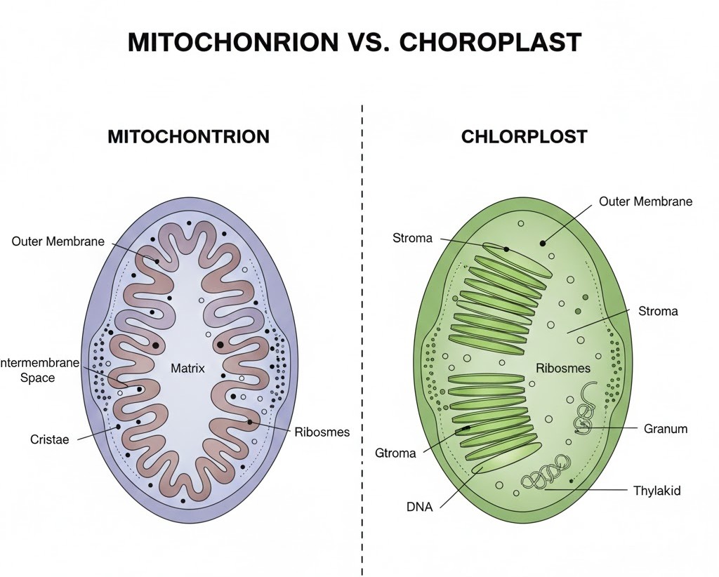
Listen up, junior! Ever wonder where your cells get the josh to finish a marathon or just survive a long coaching lecture? Meet the Mitochondria, the famous "power houses" of the cell. These sausage-shaped organelles have a double-membrane setup: a smooth outer boundary and an inner membrane thrown into folds called cristae to boost surface area for $ATP$ production. They are quite independent, like a student living in a PG, because they have their own circular DNA and $70S$ ribosomes.
NEET Checkpoint:
By what process do mitochondria divide?
(a) Mitosis
(b) Meiosis
(c) Binary fission
(d) Budding
✅ Answer: (c) Binary Fission — Simple and efficient, just like splitting a bill at a dhaba!
Next, we have Plastids, found only in plants and euglenoids. Think of them as the cell’s kitchen and pantry. Chloroplasts handle the cooking (photosynthesis), chromoplasts give fruits their bright "look at me" colours (yellow, orange, or red), and leucoplasts are the storage units.
Indian Example: Imagine your kitchen's storage jars: Amyloplasts are the sacks of rice/potatoes (starch), elaioplasts are the oil cans (fats), and aleuroplasts are the boxes of dal (proteins).
Finally, let’s talk about Ribosomes, the protein factories. These are non-membrane bound granular structures made of RNA and proteins. In eukaryotes, they are $80S$, while prokaryotes (and our "independent" organelles) use $70S$. The '$S$' stands for Svedberg’s Unit, which measures how fast they settle during a spin—basically a proxy for density and size.
Figure below:
A detailed diagram showing the double membrane of a mitochondrion with inner folds (cristae) and the matrix, contrasted with a chloroplast showing stacks of thylakoids (grana) and the stroma.
NEET Checkpoint:
What are the subunits of an $80S$ ribosome?
(a) $30S and 50S$
(b) $40S and 60S$
(c) $50S and 60S$
(d) $70S and 80S$
✅ Answer: (b) $40S$ and $60S$ — Arre, don't just add them up; biology math works differently!
NEET Checkpoint:
Where do you find chlorophyll pigments in a chloroplast?
(a) Stroma
(b) Thylakoid membranes
(c) Outer membrane
(d) Matrix
✅ Answer: (b) Thylakoid membranes — Specifically in those coin-like stacks called grana.
Practice Questions:
1. Which of the following is a colourless plastid?
(a) Chromoplast
(b) Chloroplast
(c) Leucoplast
(d) Phaeoplast
✅ Answer: (c) Leucoplast — They are the storage specialists of the cell.
2. Ribosomes are composed of:
(a) DNA and Protein
(b) RNA and Protein
(c) RNA and Lipid
(d) DNA and RNA
✅ Answer: (b) RNA and Protein — Specifically, they are ribonucleoprotein particles.
Key Takeaway
- Mitochondria: $ATP$ production, cristae, $70S$ ribosomes, circular DNA.
- Plastids: Chloroplasts (energy), Chromoplasts (colour), Leucoplasts (storage).
- Ribosomes: $80S$ (Eukaryotic) vs $70S$ (Prokaryotic), no membrane, protein synthesis.
8. Cytoskeleton & Motility
Hey junior! Ever wondered what keeps a cell from collapsing into a blob of jelly? Or how it swims around in your body fluids? Let’s clear up this funda! The cell has its own invisible "scaffolding" called the cytoskeleton. This is an elaborate network of proteinaceous structures—microtubules, microfilaments, and intermediate filaments—found in the cytoplasm. It's not just for show; it provides mechanical support, helps in motility, and maintains the shape of the cell. Think of it like the bamboo scaffolding you see on Indian construction sites—without it, the whole structure would be a mess!
NEET Checkpoint:
What are the three proteinaceous components of the cytoskeleton?
(a) Microtubules, microfilaments, intermediate filaments
(b) Actin, myosin, tubulin
(c) Collagen, elastin, keratin
(d) Centrioles, basal bodies, spindle fibers
✅ Answer: (a) Microtubules, microfilaments, and intermediate filaments — These are the structural pillars of the cell!
Now, let's talk about the cell's "engines": cilia and flagella. These are hair-like outgrowths of the cell membrane. Cilia are small and work like oars, causing movement of the cell or the surrounding fluid. Flagella are the long-distance runners—comparatively longer and responsible for cell movement.
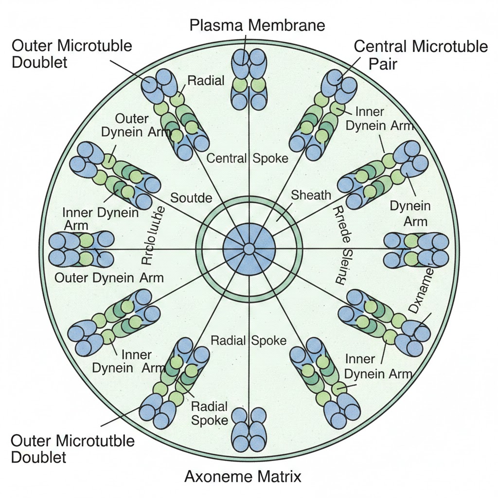
If you slice them open, you’ll find a core called the axoneme. The axoneme shows a classic $9+2$ array: nine doublets of peripheral microtubules and one pair of centrally located microtubules. The central ones are connected by bridges and enclosed by a central sheath, which is connected to the peripheral doublets by nine radial spokes. Both emerge from centriole-like structures called basal bodies. It’s like a high-tech motor inside every cell!
NEET Checkpoint:
The core of a eukaryotic cilium or flagellum is called the:
(a) Basal body
(b) Axoneme
(c) Centriole
(d) Plasma membrane
✅ Answer: (b) Axoneme — It contains the $9+2$ arrangement of microtubules.
Figure below:
A diagram showing the transverse section of a cilium/flagellum with the $9+2$ microtubule array, central sheath, and nine radial spokes.
NEET Checkpoint:
From which structure do cilia and flagella emerge?
(a) Plasma membrane
(b) Nucleus
(c) Basal bodies
(d) Golgi apparatus
✅ Answer: (c) Basal bodies — These are centriole-like structures that anchor these locomotory outgrowths.
Practice Questions:
1. The $9+2$ arrangement of microtubules is a characteristic feature of:
(a) Bacterial flagella
(b) Cytoskeleton
(c) Eukaryotic cilia and flagella
(d) Centrioles
✅ Answer: (c) — Eukaryotic cilia and flagella have this specific axonemal arrangement, while prokaryotic ones are structurally different.
2. Which function is NOT primarily associated with the cytoskeleton?
(a) Maintenance of cell shape
(b) Motility
(c) Protein synthesis
(d) Mechanical support
✅ Answer: (c) — Protein synthesis is the job of ribosomes; the cytoskeleton focuses on support, shape, and movement.
Key Takeaway
- Cytoskeleton: Network of microtubules, microfilaments, and intermediate filaments for support and shape.
- Cilia/Flagella: Locomotory outgrowths with an axoneme showing a $9+2$ array.
- Basal Bodies: The centriole-like starting point for cilia and flagella.
9. The Nucleus & Microbodies
Hey there, future doctors! Ready to dive into the "Head Office" of the cell? Just like a big Indian wedding needs a solid planner, the cell needs its nucleus to keep things running.
First described by Robert Brown in $1831$, the nucleus is where the real "funda" of life—our genetic material—is stored. The interphase nucleus (the one not currently dividing) is packed with a nucleoprotein network called chromatin, a nuclear matrix, and one or more spherical nucleoli. The nuclear envelope is a double-membrane structure with a space of $10$ to $50~nm$ between them, called the perinuclear space.
Think of nuclear pores as the high-security gates of a stadium; they are the only spots where RNA and proteins can move between the nucleus and the cytoplasm. Inside, the nucleolus is the "factory" for rhythmic RNA (rRNA) synthesis.
NEET Checkpoint:
Which scientist gave the name 'chromatin' to the material of the nucleus?
(a) Robert Brown
(b) Flemming
(c) Camillo Golgi
(d) George Palade
✅ Answer: (b) Flemming — Arre, don't confuse this with the discovery of the nucleus itself!
Now, let’s talk about the heavy lifting. Chromatin contains DNA and basic proteins called histones. Believe it or not, a single human cell has a $2~m$ long thread of DNA distributed across $46$ chromosomes. When the cell decides to divide, this chromatin condenses into structured chromosomes, each having a centromere and disc-shaped kinetochores on its sides.
NEET Checkpoint:
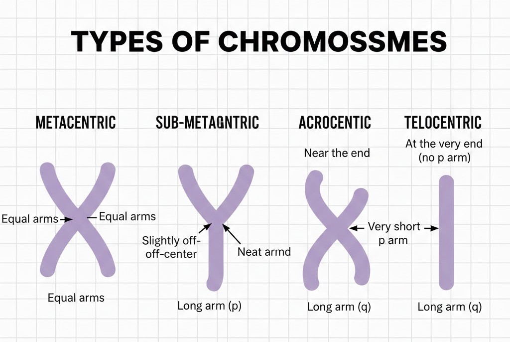
A chromosome with the centromere situated at the very end is called?
(a) Metacentric
(b) Acrocentric
(c) Telocentric
(d) Sub-metacentric
✅ Answer: (c) Telocentric — Think 'T' for Terminal!
Don't forget the Centrosome! It contains two perpendicular centrioles that look like a cartwheel. They are made of $9$ peripheral triplets of tubulin. These guys form the spindle apparatus—the "cables" that pull chromosomes apart during animal cell division. Finally, we have microbodies, which are tiny enzyme-filled vesicles found in both plant and animal cells.
NEET Checkpoint:
Where does active ribosomal RNA synthesis occur?
(a) Cytoplasm
(b) Nucleolus
(c) Centrosome
(d) Microbodies
✅ Answer: (b) Nucleolus — It's a non-membrane bound site within the nucleoplasm.
Figure bwlow:
A clear diagram showing the four types of chromosomes—metacentric, sub-metacentric, acrocentric, and telocentric—highlighting the position of the centromere and the relative length of the arms.
Practice Questions:
1. Which of the following is true about centrioles?
(a) They have a $9+2$ microtubule arrangement.
(b) They are made of 9 evenly spaced peripheral triplets of tubulin.
(c) They are found in all plant cells.
(d) They are membrane-bound organelles.
✅ Answer: (b) — Centrioles have a cartwheel-like organisation with $9$ triplets, while cilia/flagella have the $9+2$ doublet arrangement.
2. Microbodies are found in:
(a) Only animal cells
(b) Only plant cells
(c) Both plant and animal cells
(d) Only prokaryotic cells
✅ Answer: (c) — These are small vesicles containing various enzymes present in both kingdoms.
Key Takeaway
- Nucleus: Double-membraned; contains nucleolus (rRNA) and chromatin (DNA + histones).
- Chromosomes: Classified by centromere position: Metacentric (middle), Sub-metacentric (offset), Acrocentric (near end), Telocentric (terminal).
- Centrosome: Two perpendicular centrioles; 9 triplets of tubulin; forms spindle apparatus.
- Microbodies: Minute enzyme-containing vesicles in plants and animals.
10. Introduction to Biomolecules & Carbohydrates
Ever wondered what you’re actually made of, junior? To find out, we have to play a bit of a "mad scientist." We take a living tissue—like a piece of liver or a vegetable—and grind it in trichloroacetic acid ($Cl_{3}CCOOH$) using a mortar and pestle. Think of it like making a sharp, acidic chutney! Once you strain this slurry through cheesecloth, you get two fractions: the acid-soluble pool (filtrate) and the acid-insoluble fraction (retentate).
NEET Checkpoint:
Which acid is used to separate the acid-soluble and insoluble pools?
(a) $HCl$
(b) $CH_{3}COOH$
(c) $Cl_{3}CCOOH$
(d) $H_{2}SO_{4}$
✅ Answer: (c) $Cl_{3}CCOOH$ — Remember this one, yaar; it's a classic NEET favourite for chemical analysis!.
The soluble pool contains "micromolecules" with weights ranging from $18$ to $800$ daltons ($Da$). But the retentate is where the biomacromolecules live—these heavyweights, including proteins, nucleic acids, and polysaccharides, have molecular weights over $10,000$ $Da$.
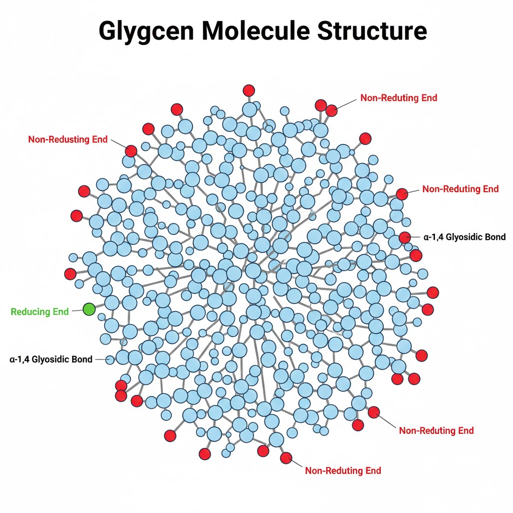
Polysaccharides are essentially long threads of sugars. For instance, cellulose is a homopolymer of glucose found in plant cell walls and even the cotton in your t-shirt. Starch (in plants) and glycogen (in animals) are your energy storehouses—like the "extra charge" in a mobile battery for a long cricket match.
NEET Checkpoint:
Which of the following is a polymer of fructose?
(a) Starch
(b) Inulin
(c) Cellulose
(d) Glycogen
✅ Answer: (b) Inulin — Don't mix this up with insulin; one is a sugar chain, the other is a hormone!.
NEET Checkpoint:
What colour is produced when starch reacts with $I_{2}$?
(a) Red
(b) Blue
(c) Yellow
(d) Green
✅ Answer: (b) Blue — Starch has a helical secondary structure that can "trap" $I_{2}$ molecules.
Figure below:
A clear diagram showing the branching structure of a glycogen molecule, highlighting the reducing and non-reducing ends.
Practice Questions:
1. The exoskeleton of a cockroach is made of which complex polysaccharide?
(a) Cellulose
(b) Chitin
(c) Glycogen
(d) Amino-sugars
✅ Answer: (b) Chitin — It's a tough homopolymer that acts like the insect's personal armour.
2. Why are lipids found in the acid-insoluble fraction despite having a weight $< 800$ $Da$?
(a) They are polymers
(b) They form vesicles
(c) They have high $O$ content
(d) They are water-soluble
✅ Answer: (b) They form vesicles — When we grind tissue, membranes break into water-insoluble vesicles that get caught in the "macro" retentate.
Key Takeaway
- Micromolecules: Acid-soluble, weight $18-800$ $Da$.
- Macromolecules: Acid-insoluble, weight $> 10,000$ $Da$ (Proteins, Nucleic Acids, Polysaccharides).
- Polysaccharides: Cellulose (structural), Starch/Glycogen (storage).
- Note: Lipids are NOT strictly macromolecules but end up in the insoluble pool.
11. Proteins & Lipids
Listen up, juniors! Today we’re diving into the "bricks and butter" of our body. First, let's talk about amino acids. Think of them as substituted methanes where the $\alpha$-carbon is the central hub. It carries four groups: a hydrogen, a carboxyl group ($-COOH$), an amino group ($-NH_{2}$), and a variable $R$ group. Depending on that $R$ group, you get 20 different types of amino acids used in proteins. For example, if $R$ is just a hydrogen, you’ve got Glycine; if it's a methyl group, it’s Alanine.
NEET Checkpoint:
Which form of amino acid exists in solutions of different pH due to its ionisable nature?
(a) Acidic
(b) Basic
(c) Zwitterionic
(d) Neutral
✅ Answer: (c) Zwitterionic — This is the form where the molecule has both positive and negative charges!.
Proteins are heteropolymers of these amino acids. Imagine a local Mumbai local train—it’s not a single type of coach; there’s general, ladies, and first class! Similarly, proteins have a sequence of different amino acids. This "positional info" is the Primary structure. Then we have Secondary (like the right-handed helix), Tertiary (folded like a hollow woolen ball), and Quaternary (multiple subunits, like the 4 subunits in human haemoglobin).
NEET Checkpoint:
Which protein is the most abundant in the entire biosphere?
(a) Collagen
(b) RuBisCO
(c) Insulin
(d) Antibody
✅ Answer: (b) RuBisCO — Don't get confused, yaaar! Collagen is most abundant in animals, but RuBisCO wins the biosphere trophy!
Finally, Lipids! These are the "oily" guys that refuse to mix with water. They include simple fatty acids like Palmitic acid ($16$ carbons) and Arachidonic acid ($20$ carbons). When fatty acids are esterified with glycerol (trihydroxy propane), we get monoglycerides or triglycerides (fats/oils). Phospholipids, like lecithin, are the stars of your cell membranes.
NEET Checkpoint:
How many carbons are present in Palmitic acid, including the carboxyl carbon?
(a) 15
(b) 16
(c) 19
(d) 20
✅ Answer: (b) 16 — Always count that carboxyl carbon, or you'll lose marks for no reason!
Practice Questions:
1. A protein is a:
(a) Homopolymer
(b) Heteropolymer
(c) Monomer
(d) Non-polymer
✅ Answer: (b) — Because it's made of different types of amino acids, not just one repeating unit.
2. Which structure is absolutely necessary for many biological activities of proteins?
(a) Primary
(b) Secondary
(c) Tertiary
(d) Quaternary
✅ Answer: (c) — Without that 3D "woolen ball" fold, the protein can't do its job!
Key Takeaway
- Proteins: Heteropolymers of 20 amino acids; structured in Primary, Secondary, Tertiary (3D), and Quaternary levels.
- Lipids: Water-insoluble; include fatty acids, glycerol-based triglycerides, and membrane phospholipids.
12. Nucleic Acids
Hey there, future doctors! Ready to decode the blueprints of life? Don't stress, yaar—it’s easier than it looks! Today we’re diving into Nucleic Acids, the "big bosses" found in the acid-insoluble fraction of living tissues. Think of them as the master scripts for everything your body does.
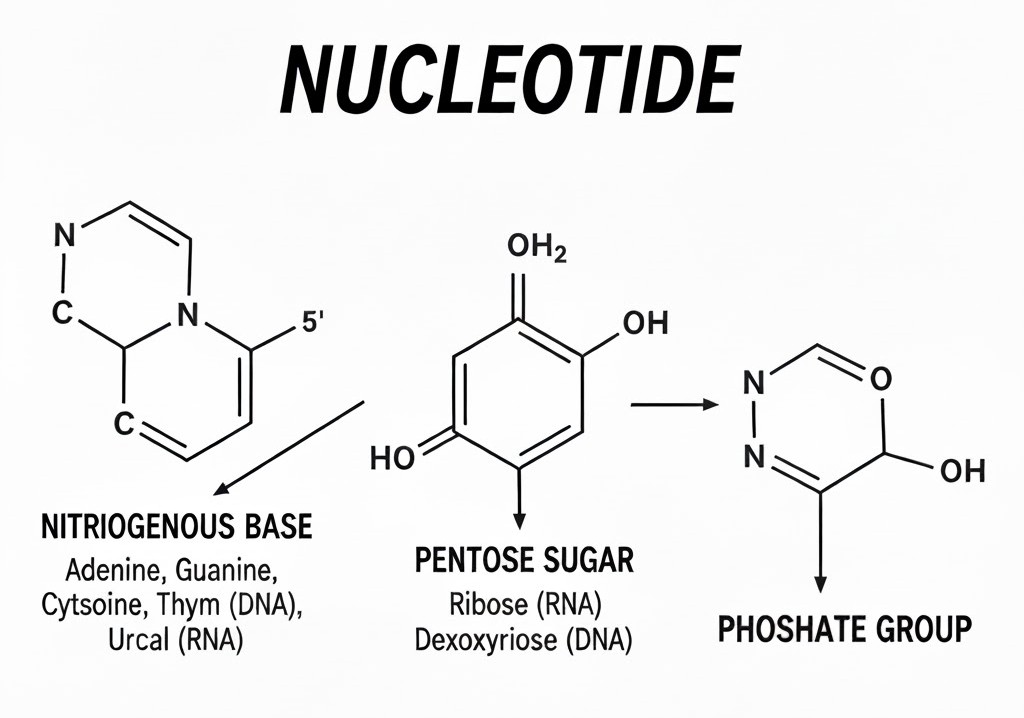
These are polynucleotides, meaning they are long chains made of building blocks called nucleotides. Each nucleotide is like a "Triple Dhamaka" combo pack containing three parts: a heterocyclic nitrogenous base, a pentose sugar, and phosphoric acid (or phosphate).
NEET Checkpoint:
What are the three chemically distinct components of a nucleotide?
(a) Sugar, protein, phosphate
(b) Sugar, nitrogenous base, phosphate
(c) Amino acid, sugar, phosphate
(d) Sugar, lipid, nitrogenous base
✅ Answer: (b) A heterocyclic compound (base), a monosaccharide (sugar), and a phosphate group — These form the basic monomeric unit.
The nitrogen bases come in two flavours: Purines (Adenine and Guanine) and Pyrimidines (Cytosine, Thymine, and Uracil). It’s like a cricket team—you’ve got your heavy-hitting purines and the agile pyrimidines.
NEET Checkpoint:
Which of these is a substituted purine?
(a) Uracil
(b) Cytosine
(c) Adenine
(d) Thymine
✅ Answer: (c) Adenine — Adenine ($A$) and Guanine ($G$) are the two main purines.
Now, the sugar makes all the difference. If the pentose sugar is ribose, it's Ribonucleic Acid (RNA). If it's 2' deoxyribose, you've got Deoxyribonucleic Acid (DNA). While both function as genetic material, DNA is the primary "hard drive" for most organisms.
Figure below:
A clear diagram showing the chemical structure of a nucleotide, highlighting the nitrogenous base, the pentose sugar (ribose/deoxyribose), and the phosphate group attached to the sugar.
NEET Checkpoint:
Name the sugar found in DNA.
(a) Glucose
(b) Ribose
(c) Deoxyribose
(d) Fructose
✅ Answer: (c) deoxyribose — This pentose sugar distinguishes DNA from RNA.
Relatable Example: Think of a local train. Each coach is a nucleotide. The passengers inside are the nitrogen bases, the floor they stand on is the sugar, and the couplings between the coaches are the phosphates. Without all three, the train (DNA) won't move!
Practice Question:
Which of the following nitrogenous bases is not found in DNA?
(a) Adenine
(b) Uracil
(c) Thymine
(d) Cytosine
✅ Answer: (b) Uracil — Uracil is specific to RNA, while DNA contains Thymine instead.
Key Takeaway
- Nucleic Acids: Polynucleotides found in the acid-insoluble pool.
- Nucleotide: Base + Sugar + Phosphate.
- Sugar Types: Ribose (RNA) and Deoxyribose (DNA).
- Bases: Purines ($A, G$) and Pyrimidines ($C, T, U$).
13. Enzymes: Classification & Action
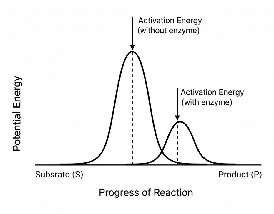
Let’s clear the funda of Enzymes—the biological magicians that make life move at lightning speed!. Almost all enzymes are proteins, though don’t forget the Ribozymes (nucleic acids acting as enzymes). Every enzyme has a special "VIP pocket" called an active site where the substrate ($S$) fits in. By forming a temporary ES complex, the enzyme lowers the activation energy, which is like reducing the height of a massive hurdle so the reaction can finish faster. Without them, your body would be slower than a rickshaw in a Mumbai traffic jam!.
NEET Checkpoint:
What happens to the rate of a reaction for every $10^{\circ}C$ change?
(a) It decreases by half
(b) It remains the same
(c) It approximately doubles
(d) It becomes zero
✅ Answer: (c) The rate doubles or decreases by half — This rule of thumb applies to both physical and chemical processes.
Enzymes are quite picky about their "working conditions." They perform best at an optimum temperature and optimum pH. If things get too hot, the protein denatures (basically breaks down), and the enzyme loses its power. Also, consider a Pani Puri stall: if there are too many customers (substrate concentration), the bhaiya can only serve so fast. Once all his hands are busy, he reaches $V_{max}$ (maximum velocity), and adding more people won’t speed things up.
NEET Checkpoint:
Which enzyme speeds up the formation of $H_{2}CO_{3}$ by 10 million times?
(a) Pepsin
(b) Carbonic anhydrase
(c) Trypsin
(d) Ligase
✅ Answer: (b) Carbonic anhydrase — It dramatically accelerates the reaction in the cytoplasm.
Enzymes are classified into six classes based on their reaction type: Oxidoreductases, Transferases, Hydrolases, Lyases, Isomerases, and Ligases. Some enzymes also need "wingmen" called co-factors to be active. If the co-factor is a permanent, tightly bound organic part (like haem), it’s a prosthetic group; if it’s a temporary visitor (like the vitamin Niacin in NAD), it’s a co-enzyme.
NEET Checkpoint:
Zinc is a co-factor for which proteolytic enzyme?
(a) Trypsin
(b) Carboxypeptidase
(c) Pepsin
(d) Chymotrypsin
✅ Answer: (b) Carboxypeptidase — Metal ions help by forming coordination bonds at the active site.
Figure below:
A graph with Potential Energy on the y-axis and Progress of Reaction on the x-axis. Show two curves: one with a high 'hump' for activation energy without an enzyme, and a much lower 'hump' for the reaction with an enzyme, leading from Substrate (S) to Product (P).
Practice Questions:
1. Competitive inhibition occurs when an inhibitor (like malonate) closely resembles:
(a) The product
(b) The enzyme
(c) The substrate
(d) The co-factor
✅ Answer: (c) The substrate — It competes for the active site because it looks similar.
2. The protein portion of an enzyme that requires a co-factor is called:
(a) Holoenzyme
(b) Apoenzyme
(c) Co-enzyme
(d) Isozyme
✅ Answer: (b) Apoenzyme — It only becomes active when the co-factor is added.
Key Takeaway
- Enzymes = Biocatalysts that lower activation energy.
- Factors: Highly sensitive to pH, Temp, and Substrate concentration.
- Classes: 6 distinct types (e.g., Hydrolases for bond breaking).
- Formula: $rate = \frac{\delta P}{\delta t}$.
14. Cell Cycle & Mitosis: The Ultimate Copy-Paste!
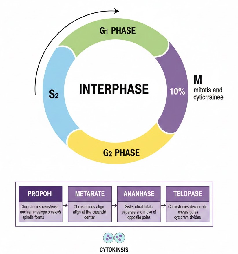
Ever wondered how a single zygote turns into a full-grown human? It’s all thanks to the cell cycle—the coordinated sequence where a cell duplicates its genome and eventually divides. Don't think of it as just division; it’s a high-stakes preparation game!
The Long Prep: Interphase
Believe it or not, cells spend more than $95\%$ of their time in Interphase, the so-called "resting phase" (though the cell is actually working hard).
* $G_{1}$ Phase: The cell is metabolically active and grows like a student during summer vacation.
* S Phase: The big one! DNA replication happens here. The DNA content doubles from $2C$ to $4C$, but the chromosome number stays the same (still $2n$).
* $G_{2}$ Phase: Proteins are synthesised as the cell does a final "viva" before division.
Indian Example: Think of Interphase like the months of planning for a big Indian wedding, while the M Phase is the actual 1-hour ceremony. All that work for one quick event!
The Big Show: Mitosis (M Phase)
Mitosis is equational division because the daughter cells are identical twins of the parent. It happens in four dramatic acts:
1. Prophase: Chromosomes condense and look like tangled woollen balls.
2. Metaphase: Chromosomes line up at the metaphase plate (equator). This is the best time to study their shape.
3. Anaphase: The "breakup" phase—centromeres split and chromatids head to opposite poles.
4. Telophase: The finish line! Nuclear envelopes reform around the chromosome clusters.
Cytokinesis then divides the cytoplasm. In animals, it’s a cell furrow (outside-in), but in plants, it’s a cell plate (inside-out) because of that rigid cell wall.
Figure below:
A clear diagram of the cell cycle showing the circular flow of G1, S, and G2 phases, followed by a detailed breakdown of the M Phase stages: Prophase, Metaphase, Anaphase, and Telophase.
NEET Checkpoint:
During which stage does DNA replication occur?
(a) G₁ phase
(b) S phase
(c) G₂ phase
(d) M phase
✅ Answer: (b) S phase — Remember, S stands for Synthesis!.
NEET Checkpoint:
Where do chromosomes align during Metaphase?
(a) At the centrosomes
(b) At the spindle poles
(c) At the equatorial/metaphase plate
(d) Randomly in the cytoplasm
✅ Answer: (c) At the equator (metaphase plate) — Perfect for a photo-op!.
NEET Checkpoint:
What is the $G_{0}$ stage?
(a) DNA synthesis phase
(b) Mitotic phase
(c) Quiescent stage
(d) Protein synthesis phase
✅ Answer: (c) Quiescent stage — Cells stay metabolically active but stop proliferating.
Practice Questions:
1. If a cell has 16 chromosomes in $G_{1}$, how many will it have after S phase?
(a) 8
(b) 16
(c) 32
(d) 64
✅ Answer: (b) 16 — DNA doubles, but chromosome number remains $2n$!.
2. Plant cytokinesis differs from animal cytokinesis due to:
(a) Centrioles
(b) Cell plate formation
(c) Furrowing
(d) Mitotic apparatus
✅ Answer: (b) Cell plate formation — Because plants have a tough cell wall!.
Key Takeaway: Mitosis Summary
- $G_{1} \rightarrow S \rightarrow G_{2}$: Prep, DNA doubling ($2C \rightarrow 4C$), and growth.
- Prophase: Condensation.
- Metaphase: Alignment at equator.
- Anaphase: Separation of chromatids.
- Telophase: Reformation of nuclei.
- Significance: Growth, cell repair, and keeping the genome intact.
15. Meiosis & Genetic Variability
Meiosis is the real deal for sexual reproduction. It’s a specialized division that takes a diploid ($2n$) cell and turns it into four haploid ($n$) gametes. Don't stress, yaar—it's easier than it looks!.
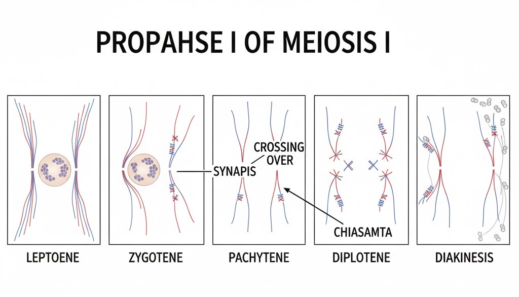
Meiosis I is the "Reduction Division" because it halves the chromosome number. The star of the show is Prophase I, which is longer and more complex than mitosis. Think of its five sub-stages like a pre-exam ritual:
1. Leptotene: Chromosomes become visible.
2. Zygotene: Homologous chromosomes pair up (synapsis) to form a bivalent or tetrad.
3. Pachytene: Here’s where the magic happens—crossing over! The enzyme recombinase swaps genetic material between non-sister chromatids.
4. Diplotene: The synaptonemal complex dissolves, leaving X-shaped chiasmata at crossing-over sites.
5. Diakinesis: Chiasmata terminalise, and the cell preps for Metaphase.
NEET Checkpoint:
During which stage does the synaptonemal complex form?
(a) Leptotene
(b) Zygotene
(c) Pachytene
(d) Diplotene
✅ Answer: (b) Zygotene — This is where chromosomes start pairing up or 'synapsing'.
Meiosis II is an "Equational Division". It resembles normal mitosis and splits the sister chromatids, resulting in four unique haploid daughter cells.
Relatable Example: Imagine two friends swapping cricket cards. After the swap (crossing over), they both have a unique set! This is why you don’t look exactly like your siblings.
NEET Checkpoint:
Which enzyme mediates crossing over?
(a) DNA polymerase
(b) Ligase
(c) Helicase
(d) Recombinase
✅ Answer: (d) Recombinase — No recombinase, no genetic mix-up, no evolution!.
Significance: Meiosis ensures the conservation of the specific chromosome number across generations. It also increases genetic variability, which is vital for evolution.
NEET Checkpoint:
What are the X-shaped structures seen in Diplotene?
(a) Centromeres
(b) Bivalents
(c) Chiasmata
(d) Synaptonemal complexes
✅ Answer: (c) Chiasmata — These are the sites where crossing over occurred.
Figure below:
A clear diagram showing the five stages of Prophase I, specifically highlighting synapsis in Zygotene, crossing over in Pachytene, and chiasmata in Diplotene.
Practice Question:
If a parental cell has $2n = 16$ chromosomes, how many chromosomes will each of the four daughter cells have after Meiosis II?
(a) 16
(b) 32
(c) 8
(d) 4
✅ Answer: (c) 8 — Meiosis reduces the chromosome number by half ($n = 8$).
Key Takeaway
- Meiosis I: Reductional division ($2n \rightarrow n$).
- Prophase I Stages: Leptotene, Zygotene (Synapsis), Pachytene (Crossing over), Diplotene (Chiasmata), Diakinesis.
- Meiosis II: Equational division resulting in 4 haploid cells.
- Significance: Chromosome conservation and genetic variation.